
Klebsiella pneumonia, also known as Friedländer pneumonia, refers to pneumonia resulting from an infection from the organism Klebsiella pneumoniae.
On this page:
Epidemiology
There tends to be a higher prevalence in older patients with alcohol use and debilitated hospitalised patients 3.
Pathology
Klebsiella pneumoniae is among the most common Gram-negative bacteria encountered by physicians worldwide and accounts for 0.5-5.0% of all cases of pneumonia 2. This organism can cause extensive pulmonary necrosis and frequent cavitation 1.
Radiographic features
Plain radiograph
It is one of the causes that could be suspected when there is cavitatory pneumonia +/- a bulging fissure sign. Often there can be extensive lobar opacification with air bronchograms.
A helpful feature that may help to distinguish from pneumococcal pneumonia is that Klebsiella pneumonia develops cavitation in 30-50% of cases (in comparison, cavitation is rare in pneumococcal pneumonia). This occurs early and progresses quickly. Massive necrosis (pulmonary gangrene) is a recognised complication.
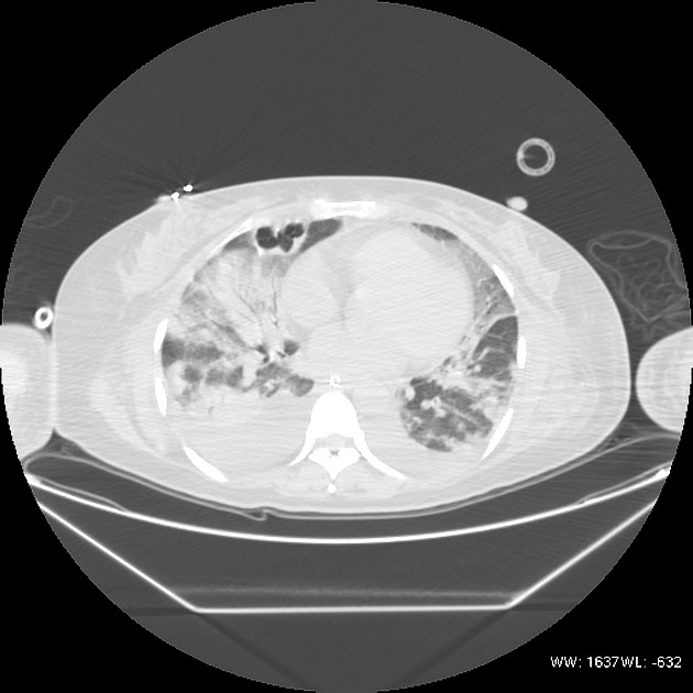
CT
There can be variable features on chest HRCT which can also depend on current other organisms. The spectrum includes
ground glass opacities: considered relatively common 5
consolidation: considered relatively common 5
intralobular reticular opacities
Necrotising pneumonia can occur as a complication 6.
History and etymology
The organism Klebsiella pneumoniae was first described by German microbiologist Carl Friedländer (1847-1887) in 1882, in a series of patients with Klebsiella pneumonia 7,8.


 Unable to process the form. Check for errors and try again.