
Left paraspinal line
Citation, DOI, disclosures and article data
Citation:
Knipe H, Hacking C, Sharma R, et al. Left paraspinal line. Reference article, Radiopaedia.org (Accessed on 23 Mar 2025) https://doi.org/10.53347/rID-23549
rID:
23549
Article created:
Disclosures:
At the time the article was created Henry Knipe had no recorded disclosures.
View Henry Knipe's current disclosures
Last revised:
Disclosures:
At the time the article was last revised Craig Hacking had no recorded disclosures.
View Craig Hacking's current disclosures
Revisions:
4 times, by
4 contributors -
see full revision history and disclosures
Systems:
Sections:
Tags:
Synonyms:
- Left paraspinous line
- Left paravertebral line
- Left paravertebral stripe
- Left paraspinous stripe
- Left paraspinal stripe
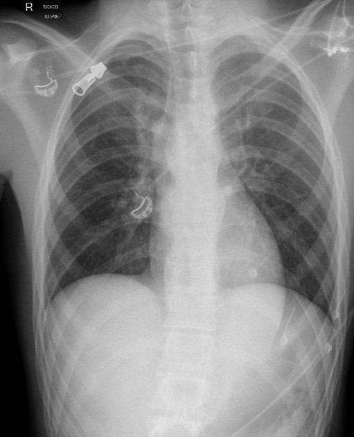
The left paraspinal (also known as the paraspinous or paravertebral) line (or stripe) is a feature of frontal chest x-rays. It is formed by the interface between the left lung and the left posterior mediastinal soft tissues 1. It is more commonly seen than the right paraspinal line.
Lateral displacement of the left paraspinal line can be due to 1-3:
- osteophytes
- mediastinal fat
- posterior mediastinal or spinal pathology
- haematoma: often from traumatic vertebral fractures
- malignancy: especially neurogenic tumours
- infection
- lymphadenopathy
- extramedullary haematopoiesis
- descending thoracic aortic aneurysm
- oesophageal varices
Radiographic features
Plain radiograph
- seen on ~35% (range 31-41%) of frontal chest x-rays 1,2
- appears as a vertical line extending from the aortic arch to (or below) the diaphragm, typically lying medial to the lateral wall of the descending thoracic aorta but occasionally will lie lateral to the aorta 1,3
References
- 1. Gibbs JM, Chandrasekhar CA, Ferguson EC et-al. Lines and stripes: where did they go?-From conventional radiography to CT. Radiographics. 2007;27 (1): 33-48. Radiographics (full text) -
- 2. Marano R, Liguori C, Savino G et-al. Cardiac silhouette findings and mediastinal lines and stripes: radiograph and CT scan correlation. Chest. 01;139 (5): 1186-96. doi:10.1378/chest.10-0660 - Pubmed citation
- 3. Whitten CR, Khan S, Munneke GJ et-al. A diagnostic approach to mediastinal abnormalities. Radiographics. 2007;27 (3): 657-71. Radiographics (full text) - doi:10.1148/rg.273065136 - Pubmed citation
Incoming Links
Related articles: Chest
- imaging techniques
-
chest radiograph
- radiography
-
approach
- ABCDE
- ABCDEFGHI
- congenital heart disease
- medical devices in the thorax
- common lines and tubes
- nasogastric tubes
- endotracheal tubes
- central venous catheters
- oesophageal temperature probe
- tracheostomy tube
- pleural catheters
- cardiac conduction devices
- prosthetic heart valve
- review areas
-
airspace opacification
- differential diagnoses of airspace opacification
- lobar consolidation
-
atelectasis
- mechanism-based
- morphology-based
- lobar lung collapse
- chest x-ray in the exam setting
- cardiomediastinal contour
- chest radiograph zones
- tracheal air column
- fissures
- normal chest x-ray appearance of the diaphragm
- nipple shadow
-
lines and stripes
- anterior junction line
- posterior junction line
- right paratracheal stripe
- left paratracheal stripe
- posterior tracheal stripe/tracheo-oesophageal stripe
- posterior wall of bronchus intermedius
- right paraspinal line
- left paraspinal line
- aortic-pulmonary stripe
- aortopulmonary window
- azygo-oesophageal recess
- spaces
- signs
- air bronchogram
- big rib sign
- Chang sign
- Chen sign
- coin lesion
- continuous diaphragm sign
- dense hilum sign
- double contour sign
- egg-on-a-string sign
- extrapleural sign
- finger in glove sign
- flat waist sign
- Fleischner sign
- ginkgo leaf sign
- Golden S sign
- Hampton hump
- haystack sign
- hilum convergence sign
- hilum overlay sign
- Hoffman-Rigler sign
- holly leaf sign
- incomplete border sign
- juxtaphrenic peak sign
- Kirklin sign
- medial stripe sign
- melting ice cube sign
- more black sign
- Naclerio V sign
- Palla sign
- pericardial fat tag sign
- Shmoo sign
- silhouette sign
- snowman sign
- spinnaker sign
- steeple sign
- straight left heart border sign
- third mogul sign
- tram-track sign
- walking man sign
- water bottle sign
- wave sign
- Westermark sign
- HRCT
-
chest radiograph
- airways
- bronchitis
- small airways disease
-
bronchiectasis
- broncho-arterial ratio
- related conditions
- differentials by distribution
- narrowing
-
tracheal stenosis
- diffuse tracheal narrowing (differential)
-
bronchial stenosis
- diffuse airway narrowing (differential)
-
tracheal stenosis
- diverticula
- pulmonary oedema
-
interstitial lung disease (ILD)
- Anti-Jo-1 antibody-positive interstitial lung disease
- drug-induced interstitial lung disease
-
hypersensitivity pneumonitis
- acute hypersensitivity pneumonitis
- subacute hypersensitivity pneumonitis
- chronic hypersensitivity pneumonitis
- aetiology
- bird fancier's lung: pigeon fancier's lung
- farmer's lung
- cheese workers' lung
- bagassosis
- mushroom worker’s lung
- malt worker’s lung
- maple bark disease
- hot tub lung
- wine maker’s lung
- woodsman’s disease
- thatched roof lung
- tobacco grower’s lung
- potato riddler’s lung
- summer-type pneumonitis
- dry rot lung
- machine operator’s lung
- humidifier lung
- shower curtain disease
- furrier’s lung
- miller’s lung
- lycoperdonosis
- saxophone lung
-
idiopathic interstitial pneumonia (mnemonic)
- acute interstitial pneumonia (AIP)
- cryptogenic organising pneumonia (COP)
- desquamative interstitial pneumonia (DIP)
- non-specific interstitial pneumonia (NSIP)
- idiopathic pleuroparenchymal fibroelastosis
- lymphoid interstitial pneumonia (LIP)
- respiratory bronchiolitis–associated interstitial lung disease (RB-ILD)
- usual interstitial pneumonia / idiopathic pulmonary fibrosis (UIP/IPF)
-
pneumoconioses
- fibrotic
- non-fibrotic
-
lung cancer
-
non-small-cell lung cancer
-
adenocarcinoma
- pre-invasive tumours
- minimally invasive tumours
- invasive tumours
- variants of invasive carcinoma
- described imaging features
- adenosquamous carcinoma
- large cell carcinoma
- primary sarcomatoid carcinoma of the lung
- squamous cell carcinoma
- salivary gland-type tumours
-
adenocarcinoma
- pulmonary neuroendocrine tumours
- preinvasive lesions
-
lung cancer invasion patterns
- tumour spread through air spaces (STAS)
- presence of non-lepidic patterns such as acinar, papillary, solid, or micropapillary
- myofibroblastic stroma associated with invasive tumour cells
- pleural invasion
- vascular invasion
- tumours by location
- benign neoplasms
- pulmonary metastases
- lung cancer screening
- lung cancer staging
-
non-small-cell lung cancer


 Unable to process the form. Check for errors and try again.