
Malakoplakia is a rare chronic granulomatous inflammatory disease that can affect any part of the body but most frequently involves the mucosal surface of the urinary bladder, causing one or more soft yellowish plaques.
On this page:
Epidemiology
Risk factors include chronic disease and immunosuppression, such as AIDS and diabetes mellitus. Malakoplakia has a peak incidence in middle age and has a reported female-to-male ratio of 4:1 which likely corresponds with the higher incidence of urinary tract infection in females.
Clinical presentation
Presenting symptoms depend on the region involved. The presence of one or more masses, sometimes with bone destruction can mimic neoplasia.
In the most common setting, when the bladder is the site of disease, patients present with gross haematuria, lower urinary tract symptoms and recurrent urinary tract infection (most commonly with Escherichia coli ). Papules, plaques and ulceration on direct visualisation during flexible cystoscopy have been described 5. Shaggy masses can mimic neoplasia.
Pathology
Defective bactericidal histiocyte function in the presence of infection leads to a chronic inflammatory response with eventual fibrosis. In the urinary tract, gram negative coliforms such as E. coli or Proteus are common inciting organisms and persist incompletely digested in phagosomes.
Location
The urinary bladder is the most frequently affected organ (40% of patients with malacoplakia). Plaques may also occur in the ureters and urethra.
Many sites of involvement have been reported:
gastrointestinal tract, most frequently rectum and colon
kidney, particularly renal transplant
genital tract
skin
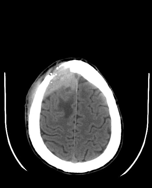
brain
tonsil
bone
adrenal
lymph node
pancreas
retroperitoneum
abdominal wall
lung
Microscopic appearance
Histology is necessary for diagnosis. Von Hansemann foamy histiocytes with intra-cytoplasmic phagocytes containing incompletely digested bacteria. During the classic middle stage, basophilic intra-cytoplasmic laminated deposits containing iron and calcium salts, called Michaelis-Gutmann bodies are pathognomonic 2,5. Special stains are helpful.
Radiographic features
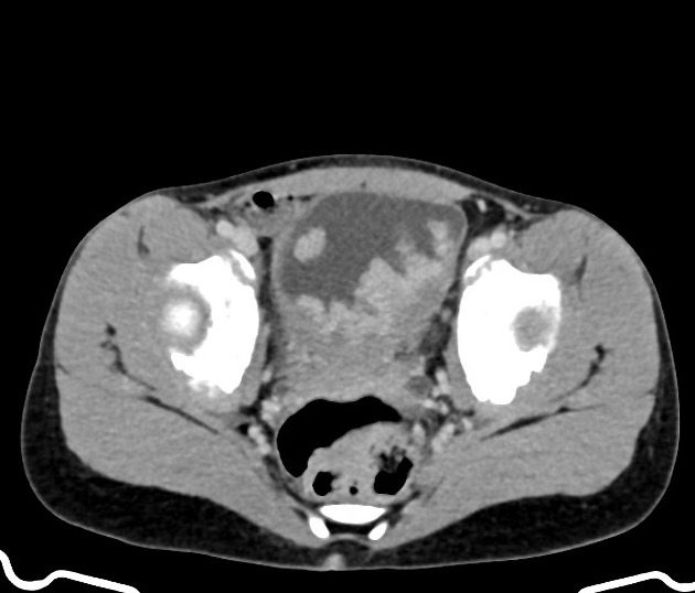
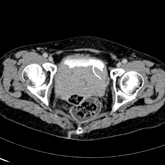
Imaging characteristics of malakoplakia are varied and depend on the region involved. In the bladder, it may present as multiple, polypoid, vascular, solid masses or as circumferential wall thickening, associated with vesicoureteral reflux or obstruction. These masses vary in size from a few millimetres to several centimetres.
Treatment and prognosis
Histological diagnosis is necessary to make the diagnosis and exclude malignancy. Although malakoplakia may be extremely aggressive, antibiotics are the mainstay of treatment. Antibiotics that concentrate in macrophages may be particularly helpful and long-term treatment may prevent recurrence. Surgery may relieve problems due to mass effect. Treatment of urinary involvement usually includes antibiotics, ascorbic acid, and a cholinergic agonist 1.
History and etymology
The term derives from "μαλακία" (malakia: "soft") and "πλακία" (plakia: "slab/plaque"). The terms "malako"plakia and "malaco"plakia are used interchangeably as latinisations of "μαλακία". The term was first used by the German pathologist David Paul von Hansemann (1858-1920) 6, but the condition was first described by L Michaelis and D Gutmann in 1902 1.
Differential diagnosis
The differential will vary greatly depending on the location of involvement.
In the urinary system, differentials include:
-
multifocal or long-segment strictures
calcification is commonly seen
-
squamous metaplasia of the urothelium
more common in bladder
-
reactive proliferative changes of the urothelium causing multiple small subepithelial cysts predominantly in the proximal ureter, urinary bladder (cystitis cystica) and the renal pelvis (pyelitis cystica)
bilaterally seen in up to 50% of cases


 Unable to process the form. Check for errors and try again.