
Metopic suture
Citation, DOI, disclosures and article data
At the time the article was created Yuranga Weerakkody had no recorded disclosures.
View Yuranga Weerakkody's current disclosuresAt the time the article was last revised Tariq Walizai had no financial relationships to ineligible companies to disclose.
View Tariq Walizai's current disclosures- Persistent metopic suture
- Frontal suture
- Median frontal suture
- Interfrontal suture
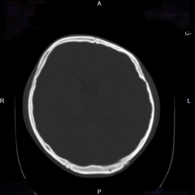
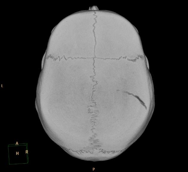
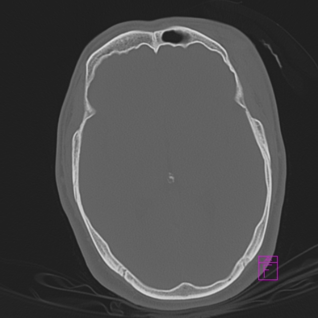
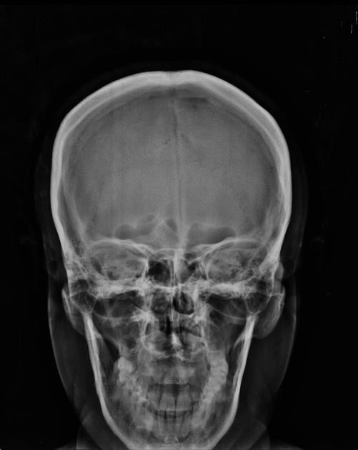
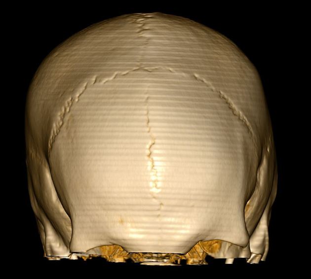
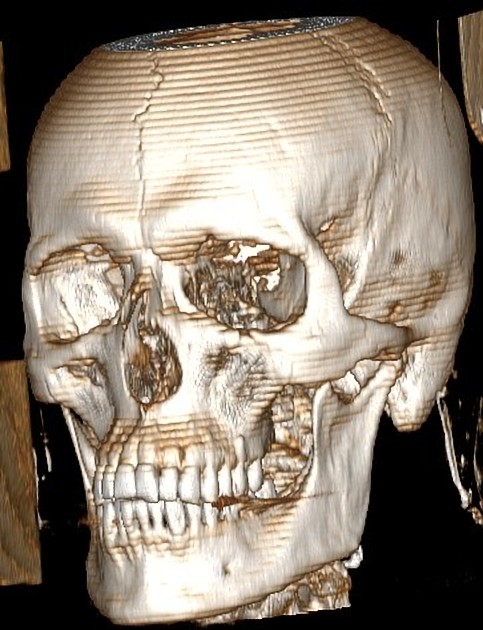
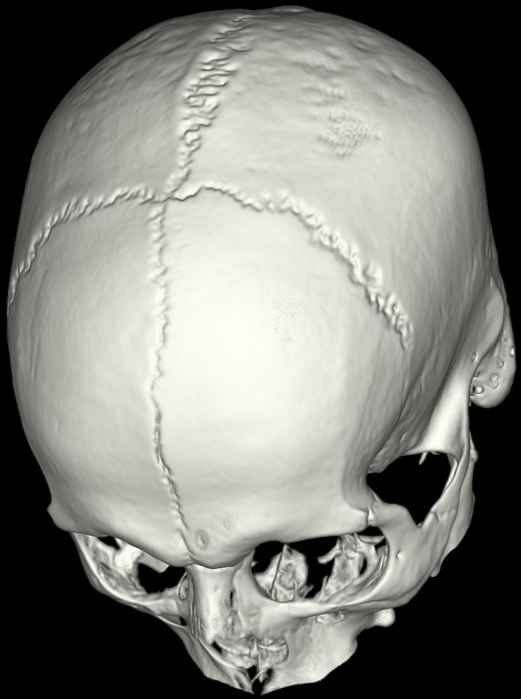
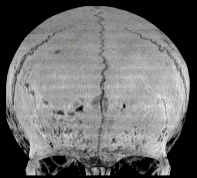
The metopic suture (also known as the frontal, interfrontal, or median frontal suture) is a vertical fibrous joint that divides the two halves of the frontal bone and is present in a newborn.
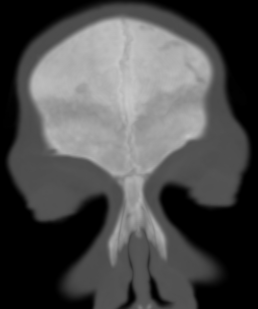
Persistent metopic sutures can be misdiagnosed as vertical skull fractures, therefore it is important to be aware of this anatomical variant.
On this page:
Gross anatomy
This suture runs through the midline across the frontal bone from the nasion to the bregma, although it may often be incomplete. It may fuse as early as 3 months of age and should fuse in nearly all patients by around 9 months of age 1-4.
Premature fusion of the suture is termed metopic synostosis (type of craniosynostosis) which can then result in trigonocephaly.
Variant anatomy
The metopic suture is usually obliterated by about 7 years of age, but in rare cases, it can persist 6 as an anatomical variant of little clinical significance but that it can be mistaken for a frontal bone fracture. Persistence of the metopic suture may be associated with frontal sinus agenesis or hypoplasia 7.
Differential diagnosis
-
metopic sutures have a characteristic midline position and demonstrate sutural interdigitations 4
References
- 1. Vu HL, Panchal J, Parker EE et-al. The timing of physiologic closure of the metopic suture: a review of 159 patients using reconstructed 3D CT scans of the craniofacial region. J Craniofac Surg. 2001;12 (6): 527-32. J Craniofac Surg (link) - Pubmed citation
- 2. Weinzweig J, Kirschner RE, Farley A et-al. Metopic synostosis: Defining the temporal sequence of normal suture fusion and differentiating it from synostosis on the basis of computed tomography images. Plast. Reconstr. Surg. 2003;112 (5): 1211-8. doi:10.1097/01.PRS.0000080729.28749.A3 - Pubmed citation
- 3. Murlimanju BV, Prabhu LV, Pai MM et-al. Median frontal sutures - incidence, morphology and their surgical, radiological importance. Turk Neurosurg. 2011;21 (4): 489-93. doi:10.5137/1019-5149.JTN .4293-11.0 - Pubmed citation
- 4. Glass RB, Fernbach SK, Norton KI et-al. The infant skull: a vault of information. Radiographics. 2004;24 (2): 507-22. Radiographics (full text) - doi:10.1148/rg.242035105 - Pubmed citation
- 5. Bademci G, Kendi T, Agalar F. Persistent metopic suture can mimic the skull fractures in the emergency setting?. Neurocirugia (Astur). 2007;18 (3): 238-40. Pubmed citation
- 6. FRCS CSS. Last's anatomy. Churchill Livingstone. ISBN:0443100330. Read it at Google Books - Find it at Amazon
- 7. Çakur B, Sumbullu MA, Durna NB. Aplasia and agenesis of the frontal sinus in Turkish individuals: a retrospective study using dental volumetric tomography. Int J Med Sci. 2011;8 (3): 278-82. Free text at pubmed - Pubmed citation
Incoming Links
- Trigonocephaly
- Persistent metopic suture with frontal sinus agensis
- Persistent metopic suture
- Persistent metopic suture
- Trigonocephaly
- Trigonocephaly
- Persistent metopic suture
- Metopic ridge
- Persistent metopic suture
- Trigonocephaly
- Trigonocephaly
- Sotos syndrome
- Persistent metopic suture
- Persistent metopic suture
- Persistent metopic suture
- Metopic suture synostosis
- Trigonocephaly
- Trigonocephaly
- Brachycephaly (bicoronal synostosis)
- Persistent metopic suture
Related articles: Anatomy: Head and neck
- skeleton of the head and neck
-
cranial vault
- scalp (mnemonic)
- fontanelle
-
sutures
- calvarial
- facial
- frontozygomatic suture
- frontomaxillary suture
- frontolacrimal suture
- frontonasal suture
- temporozygomatic suture
- zygomaticomaxillary suture
- parietotemporal suture (parietomastoid suture)
- occipitotemporal suture (occipitomastoid suture)
- sphenofrontal suture
- sphenozygomatic suture
- spheno-occipital suture (not a true suture)
- lacrimomaxillary suture
- nasomaxillary suture
- internasal suture
- basal/internal
- skull landmarks
- frontal bone
- temporal bone
- parietal bone
- occipital bone
- skull base (foramina)
-
facial bones
- midline single bones
- paired bilateral bones
- cervical spine
- hyoid bone
- laryngeal cartilages
-
cranial vault
- muscles of the head and neck
- muscles of the tongue (mnemonic)
- muscles of mastication
-
facial muscles
- epicranius muscle
- circumorbital and palpebral muscles
- nasal muscles
-
buccolabial muscles
- elevators, retractors and evertors of the upper lip
- levator labii superioris alaeque nasalis muscle
- levator labii superioris muscle
- zygomaticus major muscle
- zygomaticus minor muscle
- levator anguli oris muscle
- malaris muscle
- risorius muscle
- depressors, retractors and evertors of the lower lip
- depressor labii inferioris muscle
- depressor anguli oris muscle
- mentalis muscle
- compound sphincter
-
orbicularis oris muscle
- incisivus labii superioris muscle
- incisivus labii inferioris muscle
-
orbicularis oris muscle
- muscle of mastication
- modiolus
- elevators, retractors and evertors of the upper lip
- muscles of the middle ear
- orbital muscles
- muscles of the soft palate
- pharyngeal muscles
- suprahyoid muscles
- infrahyoid muscles
- intrinsic muscles of the larynx
- muscles of the neck
- platysma muscle
- longus colli muscle
- longus capitis muscle
- scalenus anterior muscle
- scalenus medius muscle
- scalenus posterior muscle
- scalenus pleuralis muscle
- sternocleidomastoid muscle
-
suboccipital muscles
- rectus capitis posterior major muscle
- rectus capitis posterior minor muscle
- obliquus capitis superior muscle
- obliquus capitis inferior muscle
- accessory muscles of the neck
- deep cervical fascia
-
deep spaces of the neck
- anterior cervical space
- buccal space
- carotid space
- danger space
- deep cervical fascia
- infratemporal fossa
- masticator space
- parapharyngeal space
- stylomandibular tunnel
- parotid space
- pharyngeal (superficial) mucosal space
- perivertebral space
- posterior cervical space
- pterygopalatine fossa
- retropharyngeal space
- suprasternal space (of Burns)
- visceral space
- surgical triangles of the neck
- orbit
- ear
- paranasal sinuses
- upper respiratory tract
- viscera of the neck
- blood supply of the head and neck
-
arterial supply
-
common carotid artery
- carotid body
- carotid bifurcation
- subclavian artery
- variants
-
common carotid artery
- venous drainage
-
arterial supply
- innervation of the head and neck
-
cranial nerves
- olfactory nerve (CN I)
- optic nerve (CN II)
- oculomotor nerve (CN III)
- trochlear nerve (CN IV)
-
trigeminal nerve (CN V) (mnemonic)
- trigeminal ganglion
- ophthalmic division
- maxillary division
- mandibular division
- abducens nerve (CN VI)
- facial nerve (CN VII)
-
vestibulocochlear nerve (CN VIII)
- vestibular ganglion (Scarpa's ganglion)
- glossopharyngeal nerve (CN IX)
- vagus nerve (CN X)
- (spinal) accessory nerve (CN XI)
- hypoglossal nerve (CN XII)
- parasympathetic ganglia of the head and neck
- cervical sympathetic ganglia
- greater occipital nerve
- third occipital nerve
-
cervical plexus
- muscular branches
- longus capitis
- longus colli
- scalenes
- geniohyoid
- thyrohyoid
-
ansa cervicalis
- omohyoid (superior and inferior bellies separately)
- sternothyroid
- sternohyoid
- phrenic nerve
- contribution to the accessory nerve (CN XI)
- cutaneous branches
- muscular branches
- brachial plexus
- pharyngeal plexus
-
cranial nerves
- lymphatic drainage of the head and neck
- embryological development of the head and neck


 Unable to process the form. Check for errors and try again.