
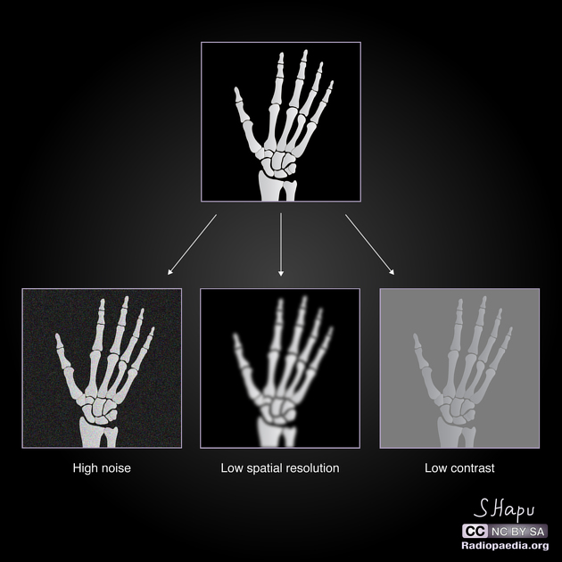
Noise, variability that is not part of a desired signal, is present in all electronic systems, and originates from a number of sources including electronic interference. It appears as an irregular granular pattern in all images and degrades image information. It may be inapparent or render images non-diagnostic, depending on the severity. Noise should not be confused with other artifacts, which are less random and should be repeatable in theory, although noise is itself an artifact.
On this page:
Quantifying noise
Image noise
Noise in an image can be quantified using the region of interest (ROI) tool placed in a region of uniform physical characteristics, like air or water. One common quantification of noise is the standard deviation in the pixel values (σ) within a region of interest with uniform physical properties. The larger the standard deviation, the worse the noise.
Radiographic issues
Plain radiograph
Noise in plain film depends on the number of discrete x-ray photons reaching the detector. The quantum noise (quantum mottle), structural noise and electronic noises are the main sources. Noise in plain radiography can be decreased by increasing the mAs which increases the number of photons reaching the detector over the duration of the exposure. If we increase the mAs by N times to the patient, noise decreases by square root N times. Note that mAs is proportional to dose.
CT
Noise (quantum noise) in CT depends on the number of discrete x-ray photons reaching the detector. See the separate article on noise (CT).
Source
The source of the electronic noise in CT is a combination of the noise of the detector system and the reconstruction kernel (sharper kernels give noisier images). Noise in a CT scan image can be decreased by increasing the mAs, by increasing the tube current or changing filters during reconstruction.
MRI
Source of noise
The main source of noise in the image is the patient's body (radiofrequency emissions due to thermal motion).The whole measurement chain of the MRI scanner (coils, electronics) also contributes to the noise.
This noise corrupts the signal coming from the transverse magnetisation variations of the intentionally excited spins (on the selected slice plane).
Reducing MRI noise
While we can never eliminate noise from imaging, the following factors can be modified to reduce it when performing MRI:
use the correct coil and ensure that it is well-tuned
use a coarse matrix
use a large field of view (FOV)
select thick slices
use narrow bandwidth
use as many NEX (number of excitations) as possible
Noise measurement
Noise in an MRI image can be quantified from the standard deviation of a homogeneous area such as the background area or from multiple acquisitions (NEX>2) 1-3.


 Unable to process the form. Check for errors and try again.