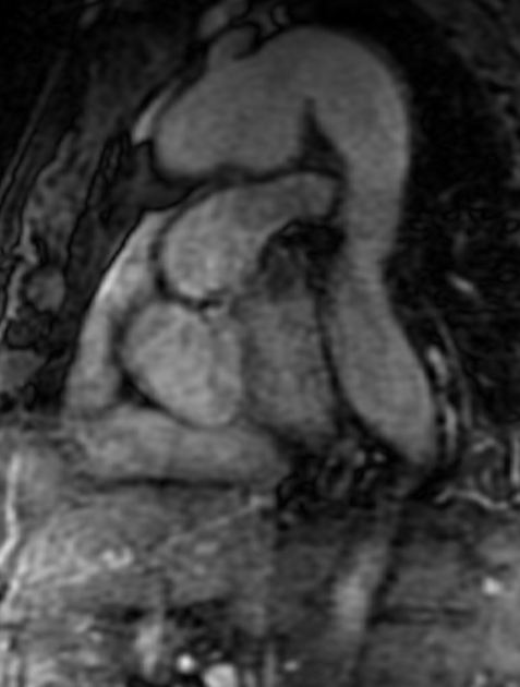
Non-contrast-enhanced MR angiography is a type of MR angiography that does not use gadolinium contrast to image the blood vessels, unlike the contrast enhanced MR angiography.
On this page:
Techniques
The earliest method that is used to image the blood vessels is by exploiting the inflow effect of the blood, also known as "time-of-flight" (TOF). The inflow effect is due to differential exposure of the stationary tissue and inflowing blood to radiofrequency (RF) excitations. Stationary tissue is repeatedly exposed to RF pulse, thus causing its longitudinal magnetization to reach approximately zero (Mz=0), producing no signal. Meanwhile, inflowing blood are not exposed to RF pulse, causing it to express full longitudinal magnetization of (Mz=1), thus producing detectable signal. The signal intensity of the flowing blood depends on blood velocity, repetition time (TR), and cross-sectional area of the blood vessel. The faster the blood velocity relative to the TR, the higher the signal of the blood. In slow-flowing blood where parts of the blood are fully saturated and the other parts are unsaturated, the signal of the blood depends upon the flip-angle and T1 relaxation time of the blood. The addition of gadolinium into the blood (as in contrast-enhanced MR angiography) will reduce the T1 relaxation time, thus increasing the signal of the slow-flowing blood 5.
Flow-compensated readout gradient is often used to correct flow-induced spin dephasing and can be applied in the readout (RO), phase encode (PE), or slice select (SS) axes 5.
Balanced steady-state free-precession gradient-echo (bSSFP) can also be used in the blood vessel imaging due to inherent flow-compensation of this sequence 5.
Types
Non-contrast-enhanced MR angiography is performed in several ways including 2:
-
flow independent
balanced steady-state free precession (bSSFP)
-
non-subtractive inflow-dependent
inflow-dependent inversion recovery (IFDIR)
quiescent interval slice-selective (QISS)
-
phase contrast angiography (PC)
2D/3D phase contrast
4D phase contrast
-
Subtractive 3D MRA
cardiac-gated 3D fast spin-echo (3D FSE or fresh blood imaging) 3
flow-sensitive dephasing (FSD)
arterial spin labeling (ASL)
Generally, these techniques are time-consuming as compared with contrast-enhanced MR angiography.
Applications
TOF is used to assess arteries of the head and neck
bSSFP is applied in imaging of the aorta and thoracic vessels
subtractive FSE and QISS can be used in peripheral arteries
IFDIR is used to image the renal arteries
PC is used in quantification of blood flow 4
