
Oesophageal leiomyoma is a benign smooth muscle neoplasm of the oesophagus. It is the most common benign tumour of the oesophagus.
On this page:
Epidemiology
It is most frequently presents in young and middle age groups (20-50 years). The overall incidence is around 8-43 per 10,000 autopsy series 4.
Clinical presentation
The clinical presentation would often depend on the size of a tumour:
- small tumours (<5 cm): usually no symptoms
- large tumours: may cause dysphagia, regurgitation, oesophageal obstruction, chest pain, cough, or bleeding (rare)
Pathology
Like other leiomyomas, they comprise smooth muscle overgrowth.
Location
They typically involve the mid-to-distal oesophagus 4.
Radiographic features
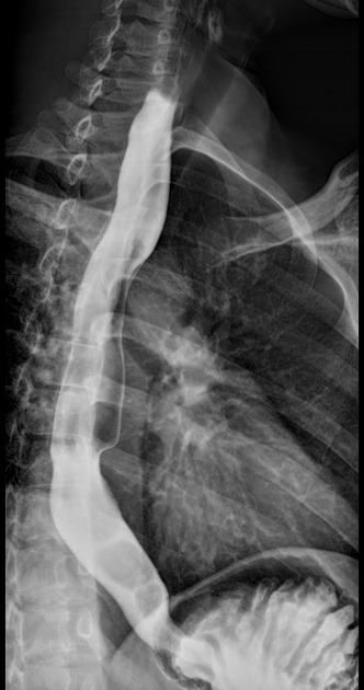
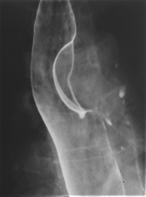
Fluoroscopy
On barium swallow, may be seen as a discrete ovoid mass that is well outlined by barium. Its borders form slightly obtuse angles with the oesophageal wall.
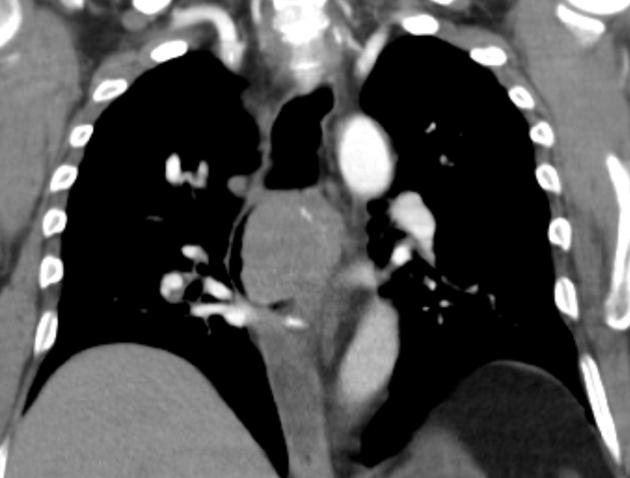
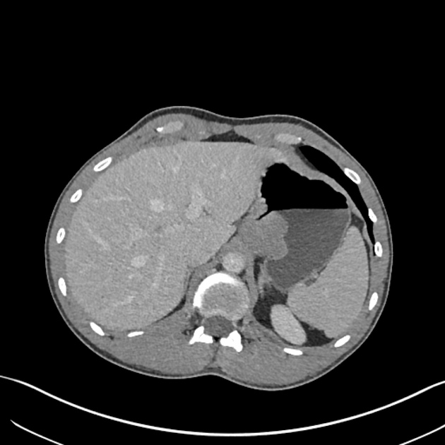
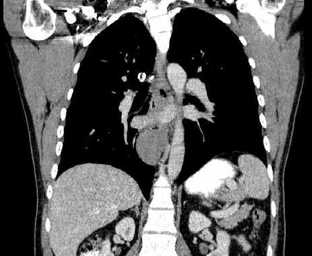
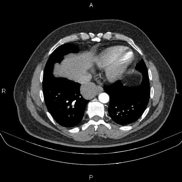
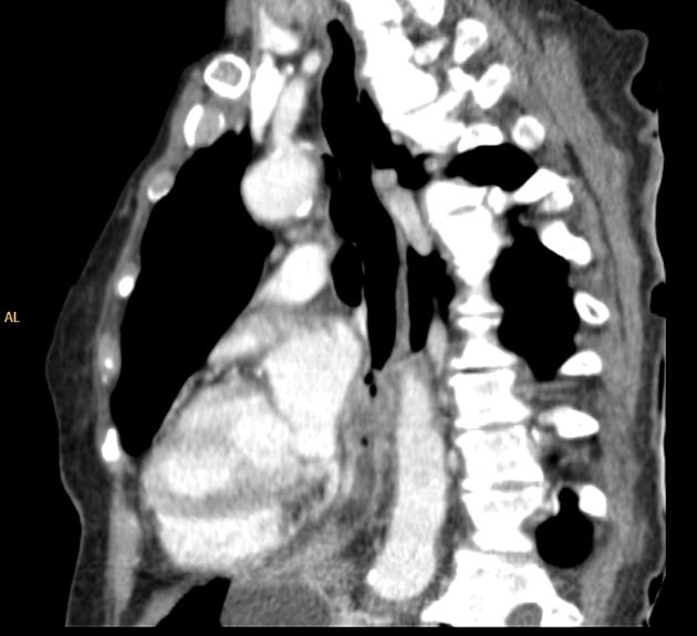
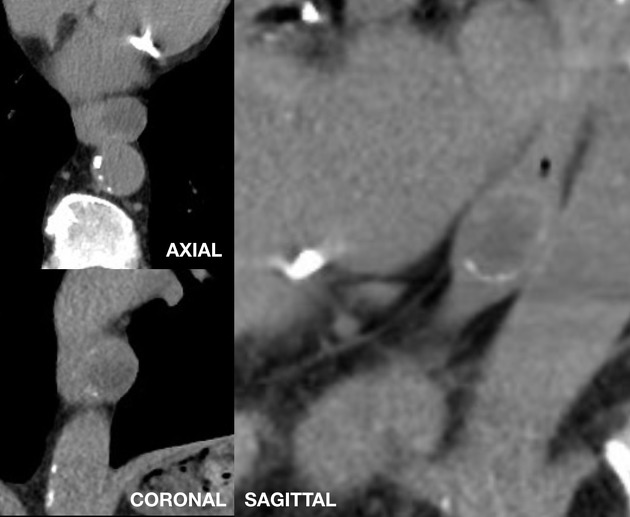
CT
Findings include:
- ovoid intramural solitary mass with a smooth surface
- the presence of calcifications is almost pathognomonic
- narrowing of the oesophageal lumen
- may displace the oesophagus
- moderate diffuse contrast-enhancement
- no signs of invasion of adjacent tissue
Treatment and prognosis
The overall prognosis of these benign tumours is excellent. If a tumour is small (<5 cm) and asymptomatic treatment is not necessary. Otherwise, surgical enucleation is recommended 1,5.
Differential diagnosis
Imaging differential considerations include:
- oesophageal GIST
- oesophageal carcinoma
- oesophageal leiomyosarcoma
- oesophageal hamartoma
- other mediastinal tumours arising close to the oesophagus
- foreign body


 Unable to process the form. Check for errors and try again.