
Os subtibiale
Citation, DOI, disclosures and article data
At the time the article was created Mohammad Taghi Niknejad had no recorded disclosures.
View Mohammad Taghi Niknejad's current disclosuresAt the time the article was last revised Ammar Ashraf had no recorded disclosures.
View Ammar Ashraf's current disclosures- Os-subtibiale
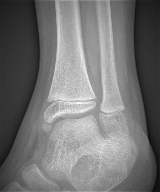
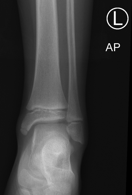
An os subtibiale is a rare, genuine accessory ossicle and normal variant related to the posterior colliculus of the medial malleolus 1. It is present in ~1% of the population 5.
On this page:
Clinical presentation
They usually are asymptomatic although they may eventually cause painful syndromes or degenerative changes in response to overuse and trauma.
Pathology
Normally, the secondary centre of ossification of the distal tibia appears during the first year of life. It fuses with the shaft at ~15 years in females and ~17 years in males. The medial malleolus is generally an extension from the distal epiphysis and ossifies in the seventh year.
An accessory bone is formed when the separate epiphysis of the medial malleolus fails to fuse with the distal tibia.
Radiographic features
Plain radiograph
- projects posteriorly, adjacent to the posterior colliculus
- typically measures more than 4 mm
- well-rounded 5
Differential diagnosis
- malleolar fracture (when radiographs are taken following an injury to the ankle) 3, 4
- ossicles related to the anterior colliculus (unfused medial malleolus ossification centre)
- present in ~2% of the population 5
- differentiated from other ossicles in this region by its smaller size to os subtibiale, its rounded and well-defined shape, and its anterior position 1,5
- post-traumatic ossification of the deltoid ligament complex 5

References
- 1. Coral A. The radiology of skeletal elements in the subtibial region: incidence and significance. Skeletal Radiol. 1987;16 (4): 298-303. Pubmed citation
- 2. Coskun N, Yuksel M, Cevener M et-al. Incidence of accessory ossicles and sesamoid bones in the feet: a radiographic study of the Turkish subjects. Surg Radiol Anat. 2009;31 (1): 19-24. Surg Radiol Anat (full text) - doi:10.1007/s00276-008-0383-9 - Pubmed citation
- 3. Park HG, Sim JA, Koh YH. Posterior tibial tendon dysfunction secondary to os subtibiale impingement: a case report. Foot Ankle Int. 2005;26 (2): 184-6. Pubmed citation
- 4. Bellapianta JM, Andrews JR, Ostrander RV. Bilateral os subtibiale and talocalcaneal coalitions in a college soccer player: a case report. J Foot Ankle Surg. 2011;50 (4): 462-5. doi:10.1053/j.jfas.2011.03.016 - Pubmed citation
- 5. Calder JDF. Sporting Injuries to the Foot & Ankle, An Issue of Foot and Ankle Clinics, (The Clinics: Orthopedics). Elsevier. ISBN:B00D9BIPHW. Read it at Google Books - Find it at Amazon
Incoming Links
Related articles: Anatomy: Lower limb
- skeleton of the lower limb
- joints of the lower limb
-
hip joint
- ligaments
- muscles
- additional structures
- hip joint capsule
- zona orbicularis
- iliotibial band
-
hip bursae
- anterior
- iliopsoas bursa (iliopectineal bursa)
- lateral
- subgluteal bursae
- greater trochanteric bursa (subgluteus maximus bursa)
- subgluteus medius bursa
- subgluteus minimus bursa
- gluteofemoral bursa
- subgluteal bursae
- postero-inferior
- anterior
- ossification centres
-
knee joint
- ligaments
- anterior cruciate ligament
- posterior cruciate ligament
- medial collateral ligament
- lateral collateral ligament
- meniscofemoral ligament (mnemonic)
-
posterolateral ligamentous complex
- arcuate ligament
- patellar tendon and quadriceps tendon
- anterolateral ligament
- posterior oblique ligament
- oblique popliteal ligament
- medial patellofemoral ligament
- additional structures
- extensor mechanism of the knee
- groove for the popliteus tendon
- knee bursae
- anterior bursae
- medial bursae
- lateral bursae
- posterior bursae
- knee capsule
- lateral patellar retinaculum
- medial patellar retinaculum
- menisci
- pes anserinus (mnemonic)
- ossification centres
- ligaments
- tibiofibular joints
-
ankle joint
- regional anatomy
- medial ankle
- lateral ankle
- anterior ankle
- ligaments
- medial collateral (deltoid) ligament
- lateral collateral ligament
- additional structures
- ankle bursae
- ossification centres of the ankle
- variants
- regional anatomy
- foot joints
- subtalar joint
- mid-tarsal (Chopart) joint
-
tarsometatarsal (Lisfranc) joint
- ligaments
- intermetatarsal joint
- metatarsophalangeal joint
- interphalangeal joint
- ossification centres
-
hip joint
- spaces of the lower limb
-
muscles of the lower limb
- muscles of the pelvic group
- muscles of the thigh
- muscles of the leg
- anterior compartment of the leg
- posterior compartments of the leg
- lateral compartment of the leg
- muscles of the foot
- dorsal muscles
- plantar muscles
- 1st layer
- 2nd layer
- 3rd layer
- 4th layer
- accessory muscles of the lower limb
- accessory gluteal muscles
-
accessory muscles of the ankle
- accessory peroneal muscles
- accessory flexor digitorum longus muscle
- accessory soleus muscle
- peroneocalcaneus internus muscle
- tibiocalcaneus internus muscle
- extensor hallucis capsularis tendon
- anterior fibulocalcaneus muscle
- accessory extensor digiti secundus muscle
- tibioastragalus anticus of Gruber muscle
- vascular supply of the lower limb
- arterial supply of the lower limb
- venous drainage of the lower limb
- innervation of the lower limb
- lymphatic system of the lower limb
- lymphatic pathways
- anteromedial group
- anterolateral group
- posteromedial group
- posterolateral group
- lower limb lymph nodes
- lymphatic pathways


 Unable to process the form. Check for errors and try again.