

Palatine bone
Citation, DOI, disclosures and article data
At the time the article was created Liam Pugh had no recorded disclosures.
View Liam Pugh's current disclosuresAt the time the article was last revised Joachim Feger had no financial relationships to ineligible companies to disclose.
View Joachim Feger's current disclosures- Os palatinum
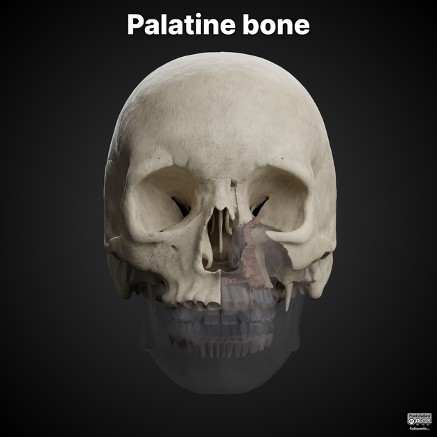
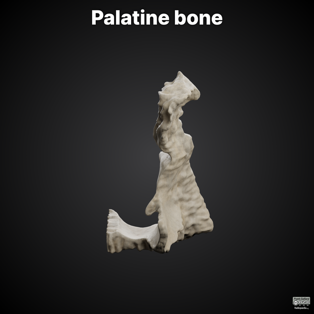
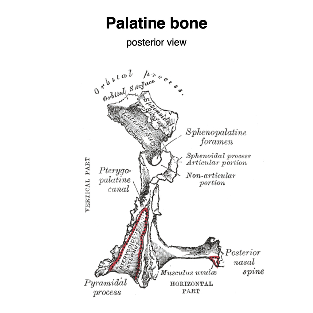
The palatine bones are paired L-shaped bones joined at the midline. They form the hard palate with the maxillary bones. They also form part of the floor of the nasal cavity (the hard palate separates the oral cavity from the nasal cavity).
Gross anatomy
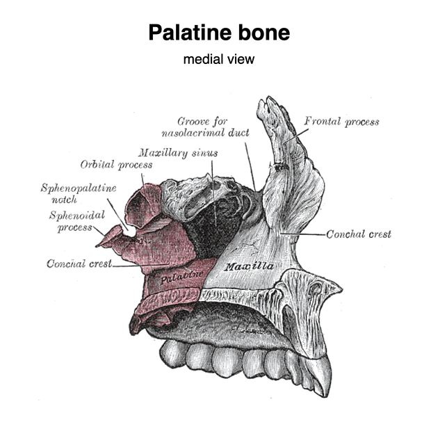
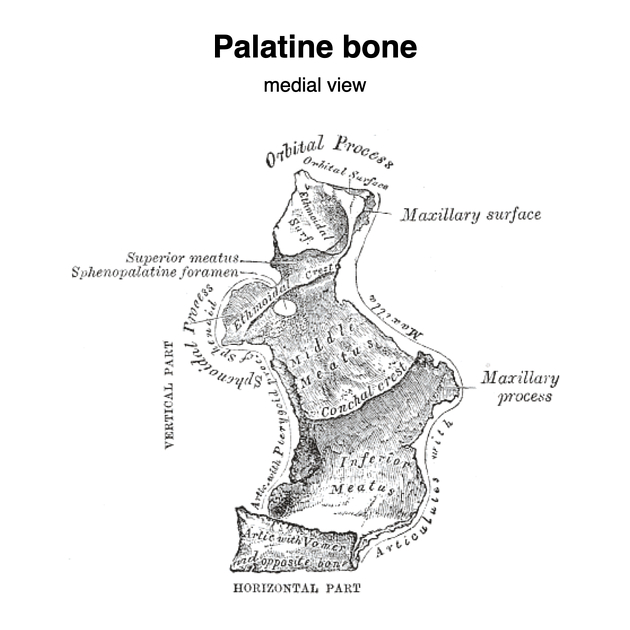
The palatine bones are located at the back of the nasal cavity, between the maxillae and the sphenoid. Each bone consists of a horizontal and perpendicular plate forming an L-shape. There are three processes, the pyramidal, orbital and sphenoidal.
They make structural contributions to the hard palate, nasal cavity, orbital floor and pterygopalatine fossa.
Articulations
with the palatine process of the maxilla anteriorly forming the transverse palatine suture
with its opposite counterpart in the midline
with the vomer
with the inferior concha
with both the body and the medial pterygoid plate of the sphenoid
with the ethmoid
Attachments
tensor palati to the posterior border of the horizontal plate
muscle of the uvula to the nasal spine of the horizontal plate
superficial head of the medial pterygoid muscle to the pyramidal process
Foramina
-
transmits the greater palatine vessels and nerve from the pterygopalatine fossa to the oral cavity
formed by the greater palatine groove and converted to a canal by articulation with the maxilla
-
transmits the lesser palatine nerves and vessels
-
formed by the sphenopalatine notch of the palatine bone articulating with the body of the sphenoid
connects the pterygopalatine fossa to the nasal cavity and transmits the sphenopalatine artery and vein, nasopalatine nerve and posterior superior nasal nerves
Ossification
intramembranous ossification appears eighth week in utero
at birth, the horizontal and perpendicular plates are of equal size
References
- 1. Susan Standring. Gray's Anatomy. (2015) ISBN: 9780702052309 - Google Books
- 2. Mcminn. Last's Anatomy. (2003) ISBN: 9780729537520 - Google Books
Incoming Links
- Lesser palatine foramina
- Maxilla
- Ethmoid bone
- Infraorbital nerve
- Inferior orbital fissure
- Lateral posterior inferior nasal nerve
- Hard palate
- Pterygopalatine fossa
- Nasal cavity
- Bony orbit
- Sphenoid bone
- Lesser palatine nerves
- Skull
- Palatovaginal canal
- Facial bones
- Lamina papyracea
- Greater palatine foramen
- Medial pterygoid muscle
- Nasal septum
- Muscle of the uvula
Related articles: Anatomy: Head and neck
- skeleton of the head and neck
-
cranial vault
- scalp (mnemonic)
- fontanelle
-
sutures
- calvarial
- facial
- frontozygomatic suture
- frontomaxillary suture
- frontolacrimal suture
- frontonasal suture
- temporozygomatic suture
- zygomaticomaxillary suture
- parietotemporal suture (parietomastoid suture)
- occipitotemporal suture (occipitomastoid suture)
- sphenofrontal suture
- sphenozygomatic suture
- spheno-occipital suture (not a true suture)
- lacrimomaxillary suture
- nasomaxillary suture
- internasal suture
- basal/internal
- skull landmarks
- frontal bone
- temporal bone
- parietal bone
- occipital bone
- skull base (foramina)
-
facial bones
- midline single bones
- paired bilateral bones
- cervical spine
- hyoid bone
- laryngeal cartilages
-
cranial vault
- muscles of the head and neck
- muscles of the tongue (mnemonic)
- muscles of mastication
-
facial muscles
- epicranius muscle
- circumorbital and palpebral muscles
- nasal muscles
-
buccolabial muscles
- elevators, retractors and evertors of the upper lip
- levator labii superioris alaeque nasalis muscle
- levator labii superioris muscle
- zygomaticus major muscle
- zygomaticus minor muscle
- levator anguli oris muscle
- malaris muscle
- risorius muscle
- depressors, retractors and evertors of the lower lip
- depressor labii inferioris muscle
- depressor anguli oris muscle
- mentalis muscle
- compound sphincter
-
orbicularis oris muscle
- incisivus labii superioris muscle
- incisivus labii inferioris muscle
-
orbicularis oris muscle
- muscle of mastication
- modiolus
- elevators, retractors and evertors of the upper lip
- muscles of the middle ear
- orbital muscles
- muscles of the soft palate
- pharyngeal muscles
- suprahyoid muscles
- infrahyoid muscles
- intrinsic muscles of the larynx
- muscles of the neck
- platysma muscle
- longus colli muscle
- longus capitis muscle
- scalenus anterior muscle
- scalenus medius muscle
- scalenus posterior muscle
- scalenus pleuralis muscle
- sternocleidomastoid muscle
-
suboccipital muscles
- rectus capitis posterior major muscle
- rectus capitis posterior minor muscle
- obliquus capitis superior muscle
- obliquus capitis inferior muscle
- accessory muscles of the neck
- deep cervical fascia
-
deep spaces of the neck
- anterior cervical space
- buccal space
- carotid space
- danger space
- deep cervical fascia
- infratemporal fossa
- masticator space
- parapharyngeal space
- stylomandibular tunnel
- parotid space
- pharyngeal (superficial) mucosal space
- perivertebral space
- posterior cervical space
- pterygopalatine fossa
- retropharyngeal space
- suprasternal space (of Burns)
- visceral space
- surgical triangles of the neck
- orbit
- ear
- paranasal sinuses
- upper respiratory tract
- viscera of the neck
- blood supply of the head and neck
-
arterial supply
-
common carotid artery
- carotid body
- carotid bifurcation
- subclavian artery
- variants
-
common carotid artery
- venous drainage
-
arterial supply
- innervation of the head and neck
-
cranial nerves
- olfactory nerve (CN I)
- optic nerve (CN II)
- oculomotor nerve (CN III)
- trochlear nerve (CN IV)
-
trigeminal nerve (CN V) (mnemonic)
- trigeminal ganglion
- ophthalmic division
- maxillary division
- mandibular division
- abducens nerve (CN VI)
- facial nerve (CN VII)
-
vestibulocochlear nerve (CN VIII)
- vestibular ganglion (Scarpa's ganglion)
- glossopharyngeal nerve (CN IX)
- vagus nerve (CN X)
- (spinal) accessory nerve (CN XI)
- hypoglossal nerve (CN XII)
- parasympathetic ganglia of the head and neck
- cervical sympathetic ganglia
- greater occipital nerve
- third occipital nerve
-
cervical plexus
- muscular branches
- longus capitis
- longus colli
- scalenes
- geniohyoid
- thyrohyoid
-
ansa cervicalis
- omohyoid (superior and inferior bellies separately)
- sternothyroid
- sternohyoid
- phrenic nerve
- contribution to the accessory nerve (CN XI)
- cutaneous branches
- muscular branches
- brachial plexus
- pharyngeal plexus
-
cranial nerves
- lymphatic drainage of the head and neck
- embryological development of the head and neck


 Unable to process the form. Check for errors and try again.