
Parotid duct
Citation, DOI, disclosures and article data
At the time the article was created Jeremy Jones had no recorded disclosures.
View Jeremy Jones's current disclosuresAt the time the article was last revised Derek Smith had no financial relationships to ineligible companies to disclose.
View Derek Smith's current disclosures- Stensen's duct
- Duct of Stensen
- Stensen duct
- Parotid ducts
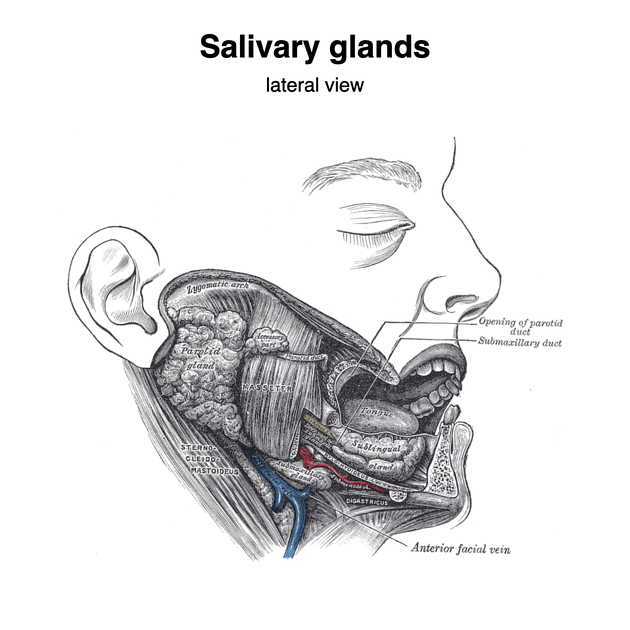
The parotid duct, also known as Stensen duct, drains saliva from the parotid gland into the oral cavity.
On this page:
Gross anatomy
Following confluence of two main tributaries within the parotid gland, the duct emerges anteriorly from its substance through the adjacent buccal fat, superficial to the masseter muscle over which it passes horizontally 9.
At the anterior border of the masseter muscle, the duct turns medially, making a right-angled turn and pierces the buccopharyngeal fascia (middle layer of the deep cervical fascia) and buccinator muscle. Here it lies in the submucosa of the oral cavity 9.
Within the submucosa, the duct travels a short distance obliquely forwards to open at the papilla, adjacent to the second maxillary molar tooth in the oral vestibule. The oblique submucosal course of the parotid duct acts as a valve, preventing reflux of contents when intraoral pressure increases 8.
Traditionally the parotid duct is described as being approximately 5 cm long and 3 mm wide 9. Studies have suggested varying diameters at different points along its length, ranging between 1.4 mm and 0.5 mm, with a maximum of 2.3 mm and a minimum of 0.1 mm, depending on the site 7.
Relations
Several other structures run alongside the parotid duct:
superiorly: transverse facial artery
inferiorly: buccal nerve
History and etymology
It is named after the Danish anatomist Niels Stensen (1638-1686) 2 (also known as Nicolaus Steno) who was the first to describe it, initially in a sheep, in 1660. His colleague Sylvius (1614-1672) confirmed its presence in the human body and van Horne in Leyden named it after Stensen 6.
Related pathology
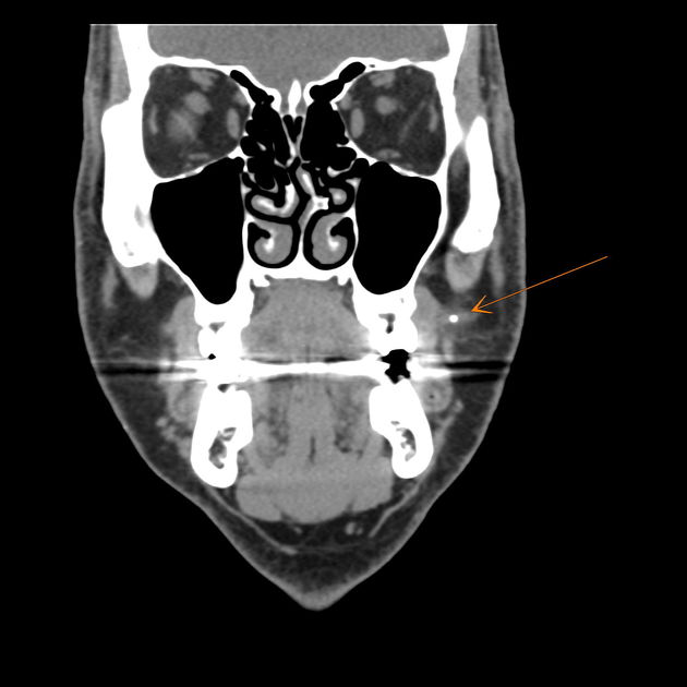
Blockage of the parotid duct can occur secondary to salivary duct stones or external compression. Either cause of obstruction can cause pain and parotitis. Stones are more common in the submandibular gland and duct.
Quiz questions

References
- 1. Lang J. Clinical anatomy of the masticatory apparatus peripharyngeal spaces. Thieme Publishing Group. ISBN:3137991013. Read it at Google Books - Find it at Amazon
- 2. Clarke E. Nicolas Stensen and the Brain [Abstract]. (1965) Proceedings of the Royal Society of Medicine. 58 (10): 749. Pubmed
- 3. Snell RS. Clinical Anatomy by Regions. Lippincott Williams & Wilkins. ISBN:160913446X. Read it at Google Books - Find it at Amazon
- 4. Moore KL, Agur AMR, Dalley AF. Clinically oriented anatomy. LWW. ISBN:1451119453. Read it at Google Books - Find it at Amazon
- 5. Last's anatomy, regional and applied. Churchill Livingstone. ISBN:044304662X. Read it at Google Books - Find it at Amazon
- 6. PORTER IH. Thomas Bartholin (1616-80) and Niels STEENSEN (1638-86). Master and pupil. (1963) Medical history. 7: 99-125. Pubmed
- 7. Zenk J, Hosemann WG, Iro H. Diameters of the main excretory ducts of the adult human submandibular and parotid gland: a histologic study. (1998) Oral surgery, oral medicine, oral pathology, oral radiology, and endodontics. 85 (5): 576-80. doi:10.1016/s1079-2104(98)90294-3 - Pubmed
- 8. Agni N. Salivary Gland Pathologies. Oral and Maxillofacial Surgery for the Clinician. 2021;:939-73. doi:10.1007/978-981-15-1346-6_46
- 9. Susan Standring. Gray's Anatomy. (2020) ISBN: 9780702077050 - Google Books
Incoming Links
- Salivary glands (Gray's illustration)
- Parotid duct stone and benign lymphoepithelial lesions
- Pleomorphic adenoma - parotid gland
- Mandible fracture
- Acute sialadenitis
- Pneumoparotid
- Buccal squamous cell carcinoma
- Parotid duct calculus
- Parotid sialolithiasis
- Sialolithiasis - parotid
- Infantile haemangioma
- Parotid duct stone
- Parotid duct alveolar rhabdomyosarcoma
- Acute left parotid sialadenitis
- Parotid sialadenitis
Related articles: Anatomy: Head and neck
- skeleton of the head and neck
-
cranial vault
- scalp (mnemonic)
- fontanelle
-
sutures
- calvarial
- facial
- frontozygomatic suture
- frontomaxillary suture
- frontolacrimal suture
- frontonasal suture
- temporozygomatic suture
- zygomaticomaxillary suture
- parietotemporal suture (parietomastoid suture)
- occipitotemporal suture (occipitomastoid suture)
- sphenofrontal suture
- sphenozygomatic suture
- spheno-occipital suture (not a true suture)
- lacrimomaxillary suture
- nasomaxillary suture
- internasal suture
- basal/internal
- skull landmarks
- frontal bone
- temporal bone
- parietal bone
- occipital bone
- skull base (foramina)
-
facial bones
- midline single bones
- paired bilateral bones
- cervical spine
- hyoid bone
- laryngeal cartilages
-
cranial vault
- muscles of the head and neck
- muscles of the tongue (mnemonic)
- muscles of mastication
-
facial muscles
- epicranius muscle
- circumorbital and palpebral muscles
- nasal muscles
-
buccolabial muscles
- elevators, retractors and evertors of the upper lip
- levator labii superioris alaeque nasalis muscle
- levator labii superioris muscle
- zygomaticus major muscle
- zygomaticus minor muscle
- levator anguli oris muscle
- malaris muscle
- risorius muscle
- depressors, retractors and evertors of the lower lip
- depressor labii inferioris muscle
- depressor anguli oris muscle
- mentalis muscle
- compound sphincter
-
orbicularis oris muscle
- incisivus labii superioris muscle
- incisivus labii inferioris muscle
-
orbicularis oris muscle
- muscle of mastication
- modiolus
- elevators, retractors and evertors of the upper lip
- muscles of the middle ear
- orbital muscles
- muscles of the soft palate
- pharyngeal muscles
- suprahyoid muscles
- infrahyoid muscles
- intrinsic muscles of the larynx
- muscles of the neck
- platysma muscle
- longus colli muscle
- longus capitis muscle
- scalenus anterior muscle
- scalenus medius muscle
- scalenus posterior muscle
- scalenus pleuralis muscle
- sternocleidomastoid muscle
-
suboccipital muscles
- rectus capitis posterior major muscle
- rectus capitis posterior minor muscle
- obliquus capitis superior muscle
- obliquus capitis inferior muscle
- accessory muscles of the neck
- deep cervical fascia
-
deep spaces of the neck
- anterior cervical space
- buccal space
- carotid space
- danger space
- deep cervical fascia
- infratemporal fossa
- masticator space
- parapharyngeal space
- stylomandibular tunnel
- parotid space
- pharyngeal (superficial) mucosal space
- perivertebral space
- posterior cervical space
- pterygopalatine fossa
- retropharyngeal space
- suprasternal space (of Burns)
- visceral space
- surgical triangles of the neck
- orbit
- ear
- paranasal sinuses
- upper respiratory tract
- viscera of the neck
- blood supply of the head and neck
-
arterial supply
-
common carotid artery
- carotid body
- carotid bifurcation
- subclavian artery
- variants
-
common carotid artery
- venous drainage
-
arterial supply
- innervation of the head and neck
-
cranial nerves
- olfactory nerve (CN I)
- optic nerve (CN II)
- oculomotor nerve (CN III)
- trochlear nerve (CN IV)
-
trigeminal nerve (CN V) (mnemonic)
- trigeminal ganglion
- ophthalmic division
- maxillary division
- mandibular division
- abducens nerve (CN VI)
- facial nerve (CN VII)
-
vestibulocochlear nerve (CN VIII)
- vestibular ganglion (Scarpa's ganglion)
- glossopharyngeal nerve (CN IX)
- vagus nerve (CN X)
- (spinal) accessory nerve (CN XI)
- hypoglossal nerve (CN XII)
- parasympathetic ganglia of the head and neck
- cervical sympathetic ganglia
- greater occipital nerve
- third occipital nerve
-
cervical plexus
- muscular branches
- longus capitis
- longus colli
- scalenes
- geniohyoid
- thyrohyoid
-
ansa cervicalis
- omohyoid (superior and inferior bellies separately)
- sternothyroid
- sternohyoid
- phrenic nerve
- contribution to the accessory nerve (CN XI)
- cutaneous branches
- muscular branches
- brachial plexus
- pharyngeal plexus
-
cranial nerves
- lymphatic drainage of the head and neck
- embryological development of the head and neck


 Unable to process the form. Check for errors and try again.