
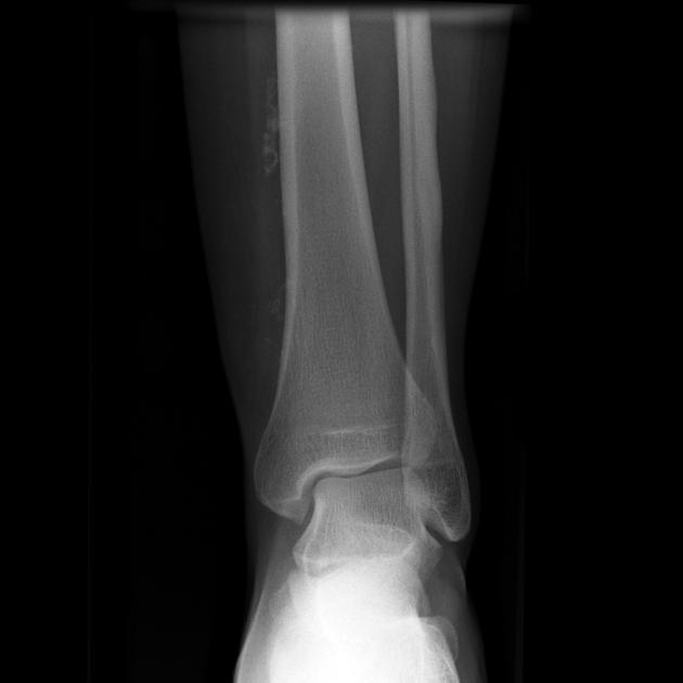
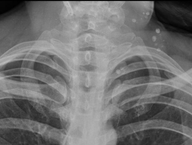
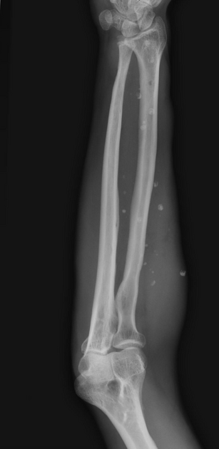
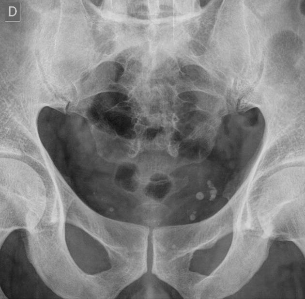
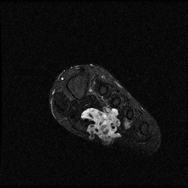
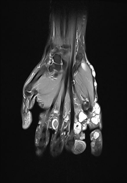
Phleboliths are literally "vein stones", and represent calcification within venous structures. They are particularly common in the pelvis, where they may mimic ureteric calculi, and are also encountered frequently in venous malformations. They can also be seen in spindle-cell haemangiomas and occasionally in macrocystic lymphatic malformations after they've bled.
On this page:
Radiographic features
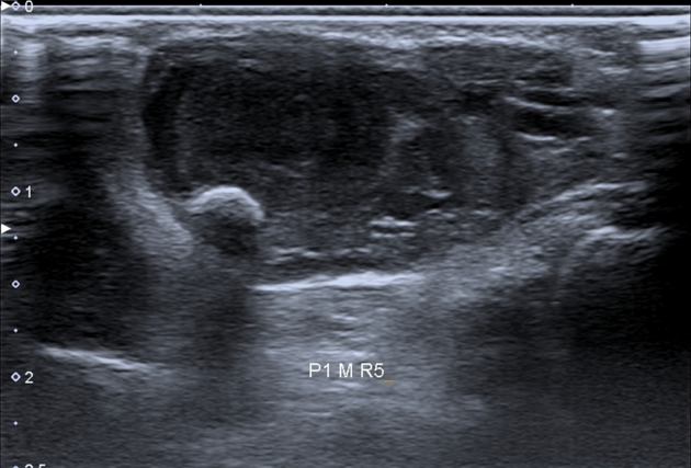
Phleboliths appear as focal calcifications, often with radiolucent centres (if present, a helpful sign to distinguish them from urolithiasis). This appearance is attributed to calcification peripherally within the vessel and is frequently seen on abdominal radiographs (66% of phleboliths 2). It can also be seen on CT provided thin sections are obtained (at 5 mm thick slices, radiolucent centres will be inapparent in 99% of phleboliths 2).
History and etymology
The word phlebolith is derived from the Ancient Greek words, φλεψ (phleps) meaning vein, and λιθος (lithos) meaning stone or rock 3.
Differential diagnosis
Two signs are helpful in distinguishing a ureteric calculus from a phlebolith:
comet tail sign: favours a phlebolith
soft tissue rim sign: favours a ureteric calculus


 Unable to process the form. Check for errors and try again.