
Presentation
Painless swelling at the foot since birth however recently suffered pain during walking and standing.
Patient Data


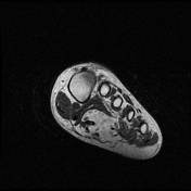
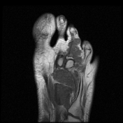
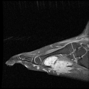
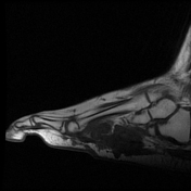
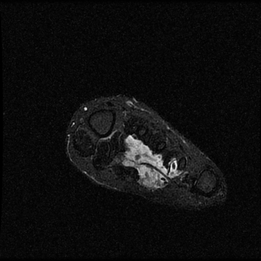
A well-defined oblong-shaped lobulated subcutaneous and deep fascial mass lesion is seen epicenter mainly along the plantar aspect of the foot as well as the 2nd, 3rd, and 4th toes. It appears isointense to the muscles on T1WI, high signal on T2WI, containing phleboliths of low signal on T2 fat sat. There is encasement of the 3rd digit from the metatarsal head to the distal phalanx as well as the proximal phalanx of the 4th digit with extension to the dorsum aspect between the 3rd and 4th interdigital spaces with subcutaneous,inter-and intramuscular extensions
Case Discussion
The MRI features are most consistent with a soft tissue hemangioma of the foot.


 Unable to process the form. Check for errors and try again.