Pisiform
Citation, DOI, disclosures and article data
At the time the article was created Aaron Wong had no recorded disclosures.
View Aaron Wong's current disclosuresAt the time the article was last revised Henry Knipe had no recorded disclosures.
View Henry Knipe's current disclosures- Pisiform bone
- Pisiforms
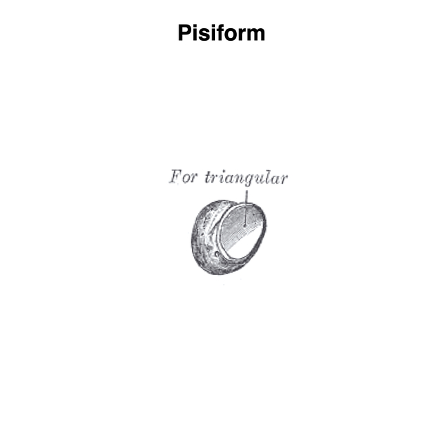
The pisiform (os pisiforme) is a small carpal bone on the medial side of the proximal carpal bones row. It is considered a sesamoid bone within the tendon flexor carpi ulnaris.
On this page:
Gross anatomy
The pisiform sits in an anterior plane to the rest of the carpal bones and articulates with the triquetrum. It has a spherical shape with a slight distolateral long axis. The articular dorsal surface is flat, forming the pisotriquetral joint, whilst the palmar surface is rough and round and provides for muscular attachment. It is the only moving structure of the carpal canal 5.
Attachments
Musculotendinous
Ligamentous
Relations
The ulnar artery sits adjacent to a lateral surface groove of the pisiform.
Arterial supply
The ulnar artery provides vascularity with branches entering both proximal and distal surfaces. Proximal vessels enter inferior to the triquetral facet near the tendon of the flexor carpi ulnaris. Superior and inferior branches run beneath the articular surface and along the palmar cortex respectively. Distal entering vessels run parallel to the palmar cortex and anastomose with the proximal vessels. Superior proximal and distal vessels form an arterial ring deep to the facet.
Variant anatomy
- os pisiforme secundarium: can be mistaken for a fracture, located at proximal pisiform pole
See accessory ossicles of the wrist.
Radiographic features
Plain radiograph
Distal avulsion and vertical fracture may occur from a direct blow, especially when the pisiform is tensed from the flexor carpi ulnaris. Mechanism often includes a fall onto an outstretched hand.
The pisotriquetral joint is a common site of wrist osteoarthritis.
Development
Often the last carpal bone to ossify, the pisiform has one ossification centre that ossifies in the ninth to twelfth year, later in males.
History and etymology
Derived from the Latin word pisum, pisiform means ‘pea-shaped’.
References
- 1. James R. Doyle. Surgical Anatomy of the Hand and Upper Extremity. (2003) ISBN: 9780397517251
- 2. Standring S, Borley N, Collins P et al. Gray's Anatomy Fortieth Edition. Churchill Livingstone. 2008.
- 3. Theumann N, Pfirrmann C, Chung C, Antonio G, Trudell D, Resnick D. Pisotriquetral Joint: Assessment with MR Imaging and MR Arthrography. Radiology. 2002;222(3):763-70. doi:10.1148/radiol.2223010466
- 4. Butler, Paul, 1952 June 4-, Mitchell, Adam W. M., Ellis, Harold, 1926-. Applied Radiological Anatomy. (1999) ISBN: 0521481104
- 5. Laude M, Le Gars D, Boudin G. [Functional Anatomy of the Pisiform Bone]. Bull Assoc Anat (Nancy). 1979;63(183):451-8. PMID 553675
Incoming Links
- Abductor digiti minimi muscle (hand)
- Hamate
- Guyon's canal
- Multicentric ossification
- Hands
- Carpal tunnel syndrome
- Accessory flexor carpi ulnaris muscle
- Carpal bones (mnemonic)
- Flexor carpi ulnaris muscle
- Palmar carpal ligament
- Flexor retinaculum (wrist)
- Carpal tunnel
- Extensor retinaculum (wrist)
- Carpal bones
- MRI of the wrist (an approach)
- Wrist series
- Bones of the upper limb
- Triquetrum
- Ossification centres of the wrist
- Isolated pisiform fracture
- Flexor carpi ulnaris - calcific tendinitis
- Pisiform fracture - flexor carpi ulnaris avulsion
- Pisiform fracture
- Isolated pisiform bone fracture (MRI)
- Flexor carpi ulnaris calcific tendinosis
- Pisiform (Gray's illustration)
- Dislocated pisiform with distal radial fracture
- Isolated pisiform fracture
- Isolated pisiform bone fracture - chronic (MRI)
- Wrist: annotated AP view
- Pisiform fracture
- Pisotriquetral joint ganglion
- Wrist - annotated carpal tunnel view
- Carpal bones - annotated x-ray
- Normal radiographic anatomy of the wrist
- Normal radiographic anatomy of the hand
- Insertion tendinopathy of flexor carpi ulnaris muscle
- Pisiform fracture
Related articles: Anatomy: Upper limb
-
skeleton of the upper limb
- clavicle
- scapula
- humerus
- radius
- ulna
- hand
- accessory ossicles of the upper limb
- accessory ossicles of the shoulder
- accessory ossicles of the elbow
-
accessory ossicles of the wrist (mnemonic)
- os centrale carpi
- os epilunate
- os epitriquetrum
- os styloideum
- os hamuli proprium
- lunula
- os triangulare
- trapezium secondarium
- os paratrapezium
- os radiostyloideum (persistent radial styloid)
- joints of the upper limb
-
pectoral girdle
-
shoulder joint
- articulations
- associated structures
- joint capsule
- bursae
- ligaments
- movements
- scapulothoracic joint
-
glenohumeral joint
- arm flexion
- arm extension
- arm abduction
- arm adduction
- arm internal rotation (medial rotation)
- arm external rotation (lateral rotation)
- circumduction
- arterial supply - scapular anastomosis
- ossification centres
-
shoulder joint
-
elbow joint
- proximal radioulnar joint
- ligaments
- associated structures
- movements
- alignment
- arterial supply - elbow anastomosis
- development
-
wrist joint
- articulations
-
ligaments
- intrinsic ligaments
- extrinsic ligaments
- radioscaphoid ligament
- dorsal intercarpal ligament
- dorsal radiotriquetral ligament
- dorsal radioulnar ligament
- volar radioulnar ligament
- radioscaphocapitate ligament
- long radiolunate ligament
- Vickers ligament
- short radiolunate ligament
- ulnolunate ligament
- ulnotriquetral ligament
- ulnocapitate ligament
- ulnar collateral ligament
- associated structures
- extensor retinaculum
- flexor retinaculum
- joint capsule
- movements
- alignment
- ossification centres
-
hand joints
- articulations
- carpometacarpal joint
-
metacarpophalangeal joints
- palmar ligament (plate)
- collateral ligament
-
interphalangeal joints
- palmar ligament (plate)
- collateral ligament
- movements
- ossification centres
- articulations
-
pectoral girdle
- spaces of the upper limb
- muscles of the upper limb
- shoulder girdle
- anterior compartment of the arm
- posterior compartment of the arm
-
anterior compartment of the forearm
- superficial
- intermediate
- deep
-
posterior compartment of the forearm (extensors)
- superficial
- deep
- muscles of the hand
-
accessory muscles
- elbow
- volar wrist midline
- palmaris longus profundus
- aberrant palmaris longus
- volar wrist radial-side
- accessory flexor digitorum superficialis indicis
- flexor indicis profundus
- flexor carpi radialis vel profundus
- accessory head of the flexor pollicis longus (Gantzer muscle, common)
- volar wrist ulnar-side
- dorsal wrist
- blood supply to the upper limb
-
arteries
- subclavian artery (mnemonic)
- axillary artery
- brachial artery (proximal portion)
- ulnar artery
- radial artery
- veins
-
arteries
- innervation of the upper limb
- intercostobrachial nerve
-
brachial plexus (mnemonic)
- branches from the roots
- branches from the trunks
- branches from the cords
- lateral cord
- posterior cord
- medial cord
- terminal branches
- lymphatic drainage of the upper limb


 Unable to process the form. Check for errors and try again.