
Planum sphenoidale
Citation, DOI, disclosures and article data
At the time the article was created Francis Deng had no recorded disclosures.
View Francis Deng's current disclosuresAt the time the article was last revised Ian Bickle had the following disclosures:
- Hexarad, I work for this out sourcing company during non NHS hours (ongoing)
These were assessed during peer review and were determined to not be relevant to the changes that were made.
View Ian Bickle's current disclosures- Jugum sphenoidale
- Sphenoidal yoke
- Jugum sphenoideum
- Sphenoid yoke
- Planum sphenoideum
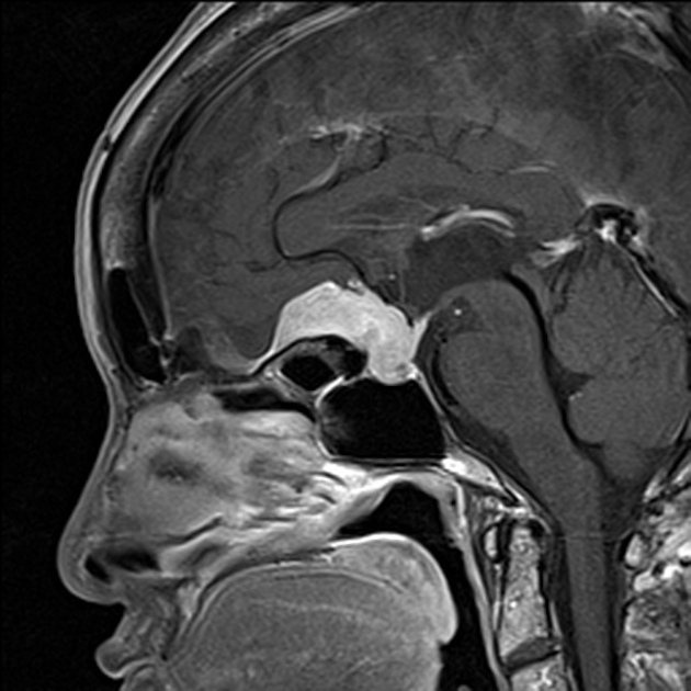
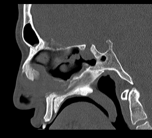
The planum sphenoidale is a common term for the anterior horizontal part of the superior surface of the body of the sphenoid bone. It is known in the Terminologia Anatomica (2019) as the jugum sphenoideum or sphenoidal yoke 1.
Gross anatomy
It forms part of the anterior skull base, separating the anterior cranial fossa from the sphenoid sinuses.
Posteriorly, the structure terminates as the limbus sphenoidale and borders the chiasmatic sulcus 2,3.
Anteriorly, the sphenoid bone articulates with the cribriform plates of the ethmoid bone 4 via the ethmoidal spine.
Laterally, the planum sphenoidale continues as the lesser wings of the sphenoid.
Related pathology
References
- 1. Chmielewski PP. New Terminologia Anatomica: Cranium and extracranial bones of head. (2019) Folia morphologica. doi:10.5603/FM.a2019.0129 - Pubmed
- 2. Bruneton JN, Drouillard JP, Sabatier JC, Elie GP, Tavernier JF. Normal variants of the sella turcica. (1979) Radiology. 131 (1): 99-104. doi:10.1148/131.1.99 - Pubmed
- 3. Kim JM, Romano A, Sanan A, van Loveren HR, Keller JT. Microsurgical anatomic features and nomenclature of the paraclinoid region. (2000) Neurosurgery. 46 (3): 670-80; discussion 680-2. doi:10.1097/00006123-200003000-00029 - Pubmed
- 4. Chong VF, Fan YF, Tng CH. Pictorial review: radiology of the sphenoid bone. (1998) Clinical radiology. 53 (12): 882-93. doi:10.1016/s0009-9260(98)80214-6 - Pubmed
Incoming Links
Related articles: Anatomy: Head and neck
- skeleton of the head and neck
-
cranial vault
- scalp (mnemonic)
- fontanelle
-
sutures
- calvarial
- facial
- frontozygomatic suture
- frontomaxillary suture
- frontolacrimal suture
- frontonasal suture
- temporozygomatic suture
- zygomaticomaxillary suture
- parietotemporal suture (parietomastoid suture)
- occipitotemporal suture (occipitomastoid suture)
- sphenofrontal suture
- sphenozygomatic suture
- spheno-occipital suture (not a true suture)
- lacrimomaxillary suture
- nasomaxillary suture
- internasal suture
- basal/internal
- skull landmarks
- frontal bone
- temporal bone
- parietal bone
- occipital bone
- skull base (foramina)
-
facial bones
- midline single bones
- paired bilateral bones
- cervical spine
- hyoid bone
- laryngeal cartilages
-
cranial vault
- muscles of the head and neck
- muscles of the tongue (mnemonic)
- muscles of mastication
-
facial muscles
- epicranius muscle
- circumorbital and palpebral muscles
- nasal muscles
-
buccolabial muscles
- elevators, retractors and evertors of the upper lip
- levator labii superioris alaeque nasalis muscle
- levator labii superioris muscle
- zygomaticus major muscle
- zygomaticus minor muscle
- levator anguli oris muscle
- malaris muscle
- risorius muscle
- depressors, retractors and evertors of the lower lip
- depressor labii inferioris muscle
- depressor anguli oris muscle
- mentalis muscle
- compound sphincter
-
orbicularis oris muscle
- incisivus labii superioris muscle
- incisivus labii inferioris muscle
-
orbicularis oris muscle
- muscle of mastication
- modiolus
- elevators, retractors and evertors of the upper lip
- muscles of the middle ear
- orbital muscles
- muscles of the soft palate
- pharyngeal muscles
- suprahyoid muscles
- infrahyoid muscles
- intrinsic muscles of the larynx
- muscles of the neck
- platysma muscle
- longus colli muscle
- longus capitis muscle
- scalenus anterior muscle
- scalenus medius muscle
- scalenus posterior muscle
- scalenus pleuralis muscle
- sternocleidomastoid muscle
-
suboccipital muscles
- rectus capitis posterior major muscle
- rectus capitis posterior minor muscle
- obliquus capitis superior muscle
- obliquus capitis inferior muscle
- accessory muscles of the neck
- deep cervical fascia
-
deep spaces of the neck
- anterior cervical space
- buccal space
- carotid space
- danger space
- deep cervical fascia
- infratemporal fossa
- masticator space
- parapharyngeal space
- stylomandibular tunnel
- parotid space
- pharyngeal (superficial) mucosal space
- perivertebral space
- posterior cervical space
- pterygopalatine fossa
- retropharyngeal space
- suprasternal space (of Burns)
- visceral space
- surgical triangles of the neck
- orbit
- ear
- paranasal sinuses
- upper respiratory tract
- viscera of the neck
- blood supply of the head and neck
-
arterial supply
-
common carotid artery
- carotid body
- carotid bifurcation
- subclavian artery
- variants
-
common carotid artery
- venous drainage
-
arterial supply
- innervation of the head and neck
-
cranial nerves
- olfactory nerve (CN I)
- optic nerve (CN II)
- oculomotor nerve (CN III)
- trochlear nerve (CN IV)
-
trigeminal nerve (CN V) (mnemonic)
- trigeminal ganglion
- ophthalmic division
- maxillary division
- mandibular division
- abducens nerve (CN VI)
- facial nerve (CN VII)
-
vestibulocochlear nerve (CN VIII)
- vestibular ganglion (Scarpa's ganglion)
- glossopharyngeal nerve (CN IX)
- vagus nerve (CN X)
- (spinal) accessory nerve (CN XI)
- hypoglossal nerve (CN XII)
- parasympathetic ganglia of the head and neck
- cervical sympathetic ganglia
- greater occipital nerve
- third occipital nerve
-
cervical plexus
- muscular branches
- longus capitis
- longus colli
- scalenes
- geniohyoid
- thyrohyoid
-
ansa cervicalis
- omohyoid (superior and inferior bellies separately)
- sternothyroid
- sternohyoid
- phrenic nerve
- contribution to the accessory nerve (CN XI)
- cutaneous branches
- muscular branches
- brachial plexus
- pharyngeal plexus
-
cranial nerves
- lymphatic drainage of the head and neck
- embryological development of the head and neck


 Unable to process the form. Check for errors and try again.