
Presentation
Visual field disturbance. CT reportedly performed elsewhere.
Patient Data
Age: 50 years
Gender: Female
From the case:
Planum sphenoidale meningioma


Download
Info

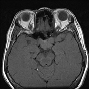
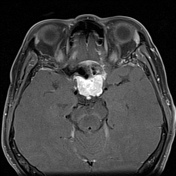
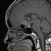
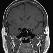
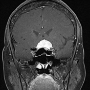
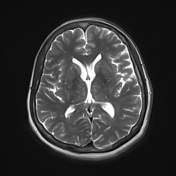
Well defined T1 isointense and T2 mildly hyperintense, homogeneously avidly enhancing mass arising from the planum sphenoidale.
A dural tail is evident.
The pituitary gland is evident beneath the mass.
Case Discussion
Meningiomas are the most common extra-axial tumors of the central nervous system. They are a non-glial neoplasm that originates from the arachnoid cap cells of the meninges and are typically benign with a low recurrence rate, but rarely can be malignant.
Meningiomas occur in a magnitude of locations, one of the less common places being the planum sphenoidale. Presentation is often due to symptoms from mass effect or incidental.


 Unable to process the form. Check for errors and try again.