
Pneumatosis cystoides intestinalis is characterised by the presence of multiple gas-filled cysts in the submucosa and/or gastrointestinal subserosa of the small intestine. It is a subtype of pneumatosis with specific features, which can occur anywhere along the gastrointestinal tract, including the colon, rectum, and mesentery. When associated with the colon it is known as pneumatosis cystoides coli. It is one of the non-surgical cause of pneumoperitoneum in neonates 14.
On this page:
Epidemiology
Pneumatosis cystoides intestinalis is an uncommon entity found in all age groups. It occurs most commonly in adults in their 5th-8th decades and has an estimated incidence of ~0.03% 11,12. This could actually be an underestimation, as most cases are asymptomatic.
Associations
About 15% of the cases have no known causes, while 85% of the cases are associated with other conditions, including 11:
COPD 13
Pathology
When the cysts rupture it may cause “benign pneumoperitoneum” as there is an absence of peritoneal irritation.
Radiographic features
Depending on the degree of gastrointestinal involvement, pneumatosis cystoides intestinalis may be diffuse, segmental, or localised.
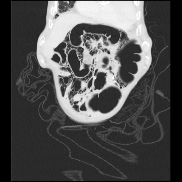
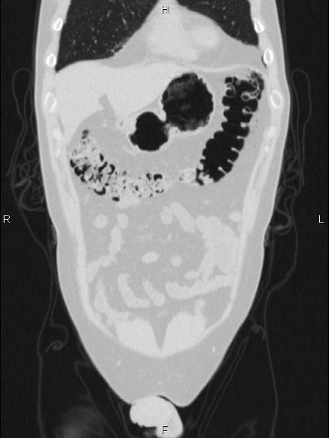
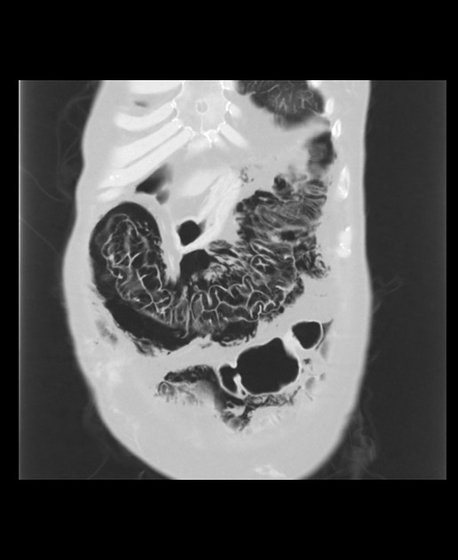
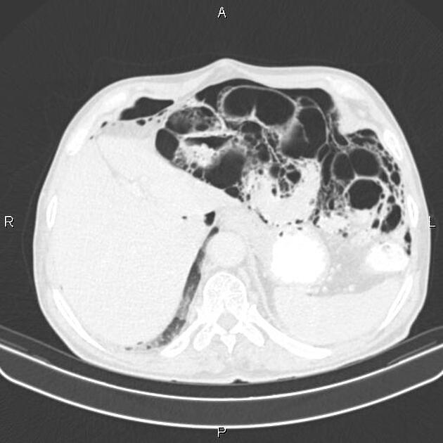
It is typically characterised by well-circumscribed cysts/”bubbles” within the walls of the hollow gastrointestinal viscera. These cysts are surrounded by regular and non-inflamed mucosa. A small volume pneumoperitoneum may be present.
If diffuse, it may appear to occupy large areas of the abdomen with a honeycomb pattern that appears similar to that of interstitial lung disease.
History and etymology
Pneumatosis cystoides intestinalis was first decribed by DuVernoi in 1783. The disease was further subcategorised by Koss in 1952 11.


 Unable to process the form. Check for errors and try again.