
The popliteus tendon is part of the popliteus musculotendinous complex together with the popliteus muscle and the popliteofibular ligament and constitutes a part of the posterolateral corner of the knee.
On this page:
Gross anatomy
The popliteus tendon ascends the posterolateral corner of the knee in a superolateral direction, running through the popliteal hiatus deep to the arcuate and fabellofibular ligaments. Before its femoral insertion, it encircles the posterolateral portion of the lateral femoral condyle beneath the lateral collateral ligament of the knee 1-3.
It is considered as an intracapsular but extrasynovial and extra-articular structure 4.
Function
The popliteus musculotendinous complex functions as a static and dynamic restraint to external rotation especially on knee flexion and as a smaller stabilizer regarding internal rotation anterior translation and varus force 1,2. It has been denoted as the “5th ligament of the knee” 3.
Attachments
The popliteus tendon inserts onto the lateral femoral condyle within the anterior portion of the popliteal sulcus 1.
Relations
The popliteus tendon serves as an origin for the popliteomeniscal fascicles. It is in close relationship to the other posterolateral corner structures of the knee including lateral collateral ligament, arcuate and fabellofibular ligaments as well as the popliteofibular ligament for which the myotendinous junction of the popliteus muscle serves as a site of origin 1. It usually forms the posterolateral border or runs through the subpopliteal recess or popliteal bursa.
Variant anatomy
In a small percentage of individuals, it is bifurcated 5.
Radiographic features
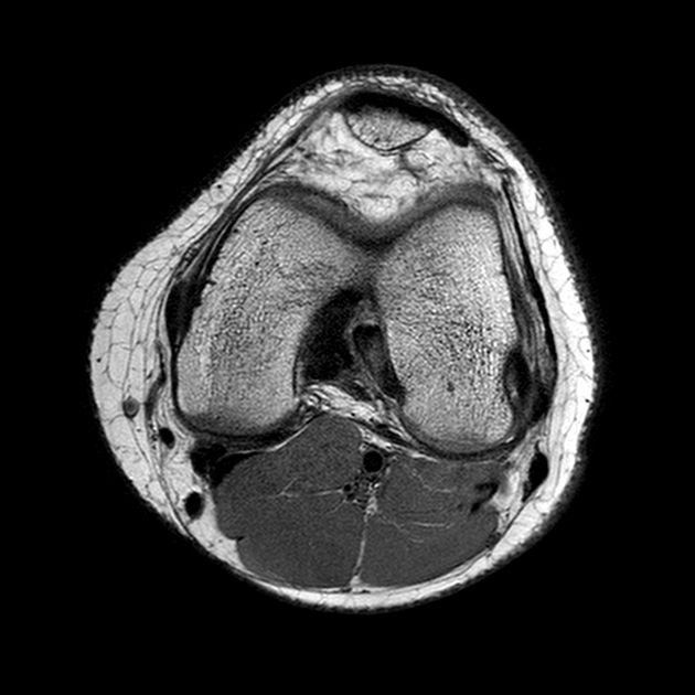
MRI
Like other tendons, the popliteus tendon is hypointense and can be easily identified on an MRI of the knee subjacent to the fibular collateral ligament.
Related pathology
Pathologies associated with the popliteus tendon include the following 6:


 Unable to process the form. Check for errors and try again.