
Pott puffy tumour refers to a non-neoplastic complication of acute sinusitis. It is characterised by a primarily subgaleal collection, subperiosteal abscess, and osteomyelitis. It is usually related to infective pathologies of the frontal sinus.
On this page:
Epidemiology
Although it may affect patients of any age, the incidence is higher in adolescence. It has become unusual since the availability of antibiotics.
Pathology
Pott puffy tumour is usually related to infection of the frontal sinus but is sometimes secondary to mastoid pathology. Rarer aetiologies include trauma, intranasal cocaine, and methamphetamine abuse, and craniotomy.
The infection erodes through the wall of the obstructed infected sinus to form a subperiosteal abscess. As expected it can be associated with extension intracranially with epidural abscess, subdural empyema, meningitis, and cerebral abscess formation. Dural sinus thrombosis is another possible complication.
Microbiology
The causative agent usually reflects the type of bacterial species responsible for community-acquired chronic sinusitis. The most common infective agents implicated are:
Streptococcus spp.
Haemophilus influenzae
Staphylococcus spp.
Klebsiella sp.
Other reported organisms include
Pasteurella multocida 6
Radiographic features
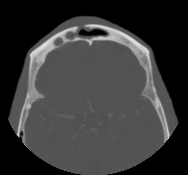
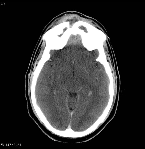
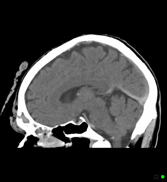
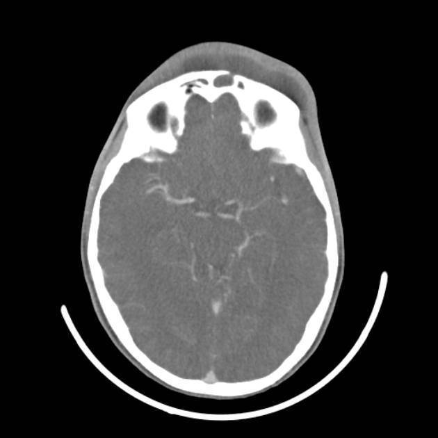
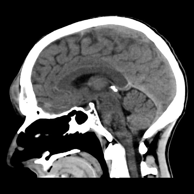
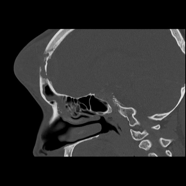
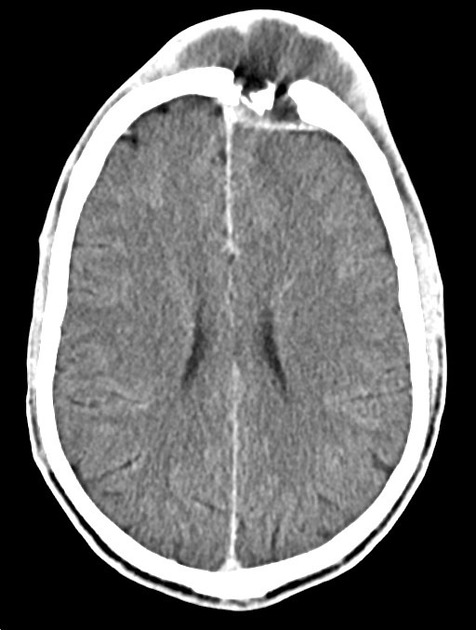
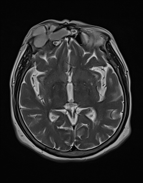
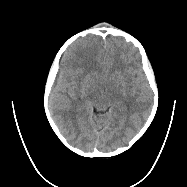
CT
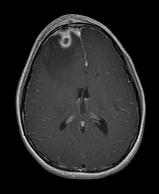
CT typically demonstrates an opacified frontal sinus with stranding and swelling of the overlying scalp. Bone algorithm will often demonstrate a defect in the anterior wall of the sinus.
Contrast may demonstrate a focal abscess, and may also allow intracranial complications to be better delineated.
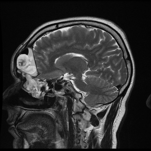
MRI
Subtle intracranial involvement is more easily seen at MR imaging. With the injection of gadolinium-based contrast material, one may see early linear enhancement of the dura mater, an extra-axial fluid collection, or an area of cerebritis or focal cerebral abscess formation.
In the scalp, peripheral or rim contrast enhancement may be seen when an organised fluid collection is present.
Treatment and prognosis
Treatment is typically surgical with drainage of the abscess and at least 6 weeks of intravenous antibiotics.
History and etymology
It was first described by Sir Percivall Pott (see Pott disease) in 1760. The characteristic forehead swelling as a result of the subgaleal collection explains the "puffy tumour" part of the name.
Differential diagnosis
Possible differential considerations on imaging grounds include:
unilateral non-Hodgkin lymphoma 10
destructive metastasis to frontal sinus 9


 Unable to process the form. Check for errors and try again.