
Proximal radioulnar joint
Citation, DOI, disclosures and article data
At the time the article was created Michael Stewart had no recorded disclosures.
View Michael Stewart's current disclosuresAt the time the article was last revised Joachim Feger had no financial relationships to ineligible companies to disclose.
View Joachim Feger's current disclosures- Proximal radioulnar joint (PRUJ)
- PRUJ
- Proximal radio-ulnar joint
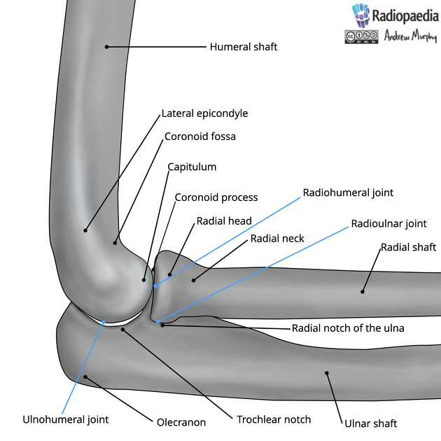
The proximal radioulnar joint is a complex pivot-type synovial joint between the circumference of the head of the radius and the ring formed by the radial notch of the ulna and the annular ligament.
Gross anatomy
The proximal radiohumeral joint is comprised of three joints
radiohumeral joint: The radial head with the capitellum
radioulnar: Radial notch with the ulnar.
ulnohumeral: the trochlea with the coronoid
Joint capsule
The joint capsule encloses the joint; both it and the synovial membrane are continuous with that of the elbow joint. The capsule is relatively thin compared to other capsules, with its anterior component anteriorly attaching the medial epicondyle and ulnar coranoid process. The posterior aspect of the capsule is thinner than its anterior component.
Movement
Rotatory movements of the head of the radius within the collar formed by the annular ligament and the radial notch, allowing supination (primarily by the action of the biceps brachii and supinator muscles) and pronation (by the action of pronator teres and pronator quadratus muscles).
Ligaments
annular ligament: strong ligament attached to the anterior and posterior aspects of the radial notch of the ulna. It (along with the radial notch of the ulna) encircles the head of the radius.
Bursa
Olecranon bursa which lies between the capsule and the triceps insertion
Blood supply
From the radial portion of the peri-articular arterial anastomosis of the elbow joint.
Innervation
From articular branches of the musculocutaneous, ulnar, median, and radial nerves.
Variants
synovial folds
-
accessory ossicles:
References
- 1. Chummy S. Sinnatamby. Last's Anatomy. (2011) ISBN: 9780702033940 - Google Books
- 2. Richard Drake, A. Wayne Vogl, Adam W. M. Mitchell. Gray's Anatomy for Students. (2023) ISBN: 9780323934237 - Google Books
Incoming Links
Related articles: Anatomy: Upper limb
-
skeleton of the upper limb
- clavicle
- scapula
- humerus
- radius
- ulna
- hand
- accessory ossicles of the upper limb
- accessory ossicles of the shoulder
- accessory ossicles of the elbow
-
accessory ossicles of the wrist (mnemonic)
- os centrale carpi
- os epilunate
- os epitriquetrum
- os styloideum
- os hamuli proprium
- lunula
- os triangulare
- trapezium secondarium
- os paratrapezium
- os radiostyloideum (persistent radial styloid)
- joints of the upper limb
-
pectoral girdle
-
shoulder joint
- articulations
- associated structures
- joint capsule
- bursae
- ligaments
- movements
- scapulothoracic joint
-
glenohumeral joint
- arm flexion
- arm extension
- arm abduction
- arm adduction
- arm internal rotation (medial rotation)
- arm external rotation (lateral rotation)
- circumduction
- arterial supply - scapular anastomosis
- ossification centres
-
shoulder joint
-
elbow joint
- proximal radioulnar joint
- ligaments
- associated structures
- movements
- alignment
- arterial supply - elbow anastomosis
- development
-
wrist joint
- articulations
-
ligaments
- intrinsic ligaments
- extrinsic ligaments
- radioscaphoid ligament
- dorsal intercarpal ligament
- dorsal radiotriquetral ligament
- dorsal radioulnar ligament
- volar radioulnar ligament
- radioscaphocapitate ligament
- long radiolunate ligament
- Vickers ligament
- short radiolunate ligament
- ulnolunate ligament
- ulnotriquetral ligament
- ulnocapitate ligament
- ulnar collateral ligament
- associated structures
- extensor retinaculum
- flexor retinaculum
- joint capsule
- movements
- alignment
- ossification centres
-
hand joints
- articulations
- carpometacarpal joint
-
metacarpophalangeal joints
- palmar ligament (plate)
- collateral ligament
-
interphalangeal joints
- palmar ligament (plate)
- collateral ligament
- movements
- ossification centres
- articulations
-
pectoral girdle
- spaces of the upper limb
- muscles of the upper limb
- shoulder girdle
- anterior compartment of the arm
- posterior compartment of the arm
-
anterior compartment of the forearm
- superficial
- intermediate
- deep
-
posterior compartment of the forearm (extensors)
- superficial
- deep
- muscles of the hand
-
accessory muscles
- elbow
- volar wrist midline
- palmaris longus profundus
- aberrant palmaris longus
- volar wrist radial-side
- accessory flexor digitorum superficialis indicis
- flexor indicis profundus
- flexor carpi radialis vel profundus
- accessory head of the flexor pollicis longus (Gantzer muscle, common)
- volar wrist ulnar-side
- dorsal wrist
- blood supply to the upper limb
-
arteries
- subclavian artery (mnemonic)
- axillary artery
- brachial artery (proximal portion)
- ulnar artery
- radial artery
- veins
-
arteries
- innervation of the upper limb
- intercostobrachial nerve
-
brachial plexus (mnemonic)
- branches from the roots
- branches from the trunks
- branches from the cords
- lateral cord
- posterior cord
- medial cord
- terminal branches
- lymphatic drainage of the upper limb


 Unable to process the form. Check for errors and try again.