
Pulmonary vein thrombosis is a rare but potentially serious condition with a number of underlying possible aetiologies.
On this page:
Clinical presentation
Often the signs and symptoms are non-specific and can range from acute (pulmonary infarction) to more insidious (progressive or recurrent pulmonary oedema). Patients may have dyspnoea, haemoptysis, chest pain, fever, and/or hypoxaemia.
Pathology
Aetiology
Causes include:
intrapulmonary neoplasm: considered most frequent cause 1
as a complication of lung transplantation or lobectomy 6
as a complication of radiofrequency ablation
mitral stenosis with a left atrial clot
hypercoagulable state
idiopathic
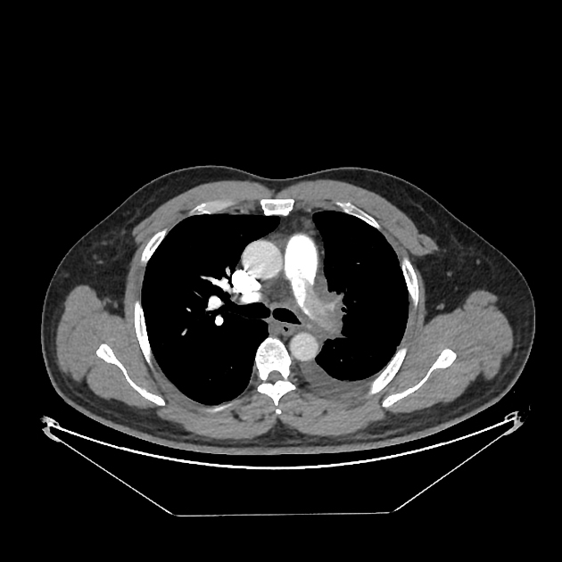
Radiographic features
CT
Chronic pulmonary vein stenosis or obstruction may demonstrate 9:
interlobular septal thickening and ground glass opacification due to localised pulmonary oedema
enlarged lymph nodes, lymphangiectasis and pleural effusion due to impaired venous drainage
venous collateral pathways - venous varices
thickening of bronchovascular bundles due to bronchovascular ectasis and remodelling of the bronchial arteries and veins
pleural plaques may be seen due to organisation of the fibrinous exudates
interstitial fibrosis may also be seen due to the accumulation of siderophages in the interstitial spaces
Treatment and prognosis
Anticoagulation use is thought to be doubtful in terms of clinical outcome but may avoid clot development in the left atrium and possibly promote recanalisation of the affected pulmonary vein 3.
Differential diagnosis
pulmonary vein flow artifact - pulmonary vein smoke


 Unable to process the form. Check for errors and try again.