
The satellite sign is a radiological sign seen on non-contrast CT of the brain in the setting of intracerebral haemorrhage, and refers to a small haemorrhage adjacent to, and separate from, the main haematoma. It is a predictor of haemorrhage expansion.
On this page:
Epidemiology
The satellite sign can be seen in ~40% of patients with intracerebral haemorrhage scanned within 6 hours of symptom onset 1-3. There is an association with hypertension and intraventricular haemorrhage 1.
Pathology
Following a haemorrhagic insult, the tissues adjacent to the haematoma are thought to develop cytotoxic oedema, eventually resulting in local ischaemia and reperfusion injury. Subsequent disruption of the blood-brain barrier then manifests as the satellite sign 1.
Radiographic features
CT
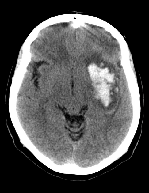
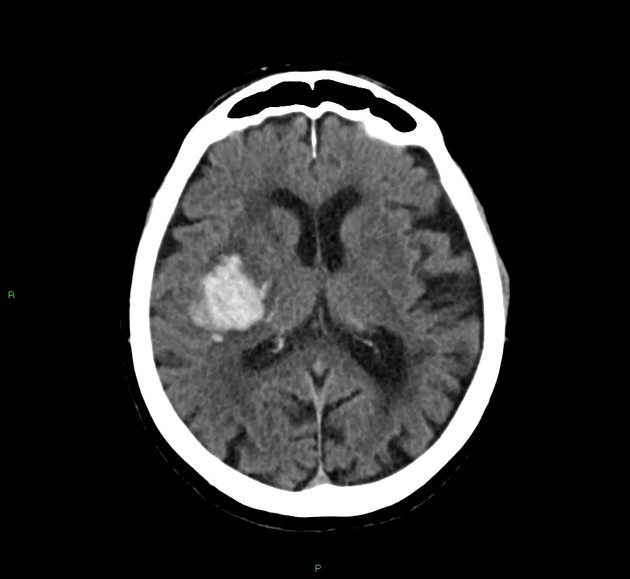
The satellite sign is defined as a small intraparenchymal hyperdense haemorrhagic focus that is clearly separated from the main haematoma on at least one CT slice. This focus can exhibit any morphology but has to 4:
measure no more than 10 mm in maximum diameter
be separated from the main haematoma by no more than 20 mm
remain intraparenchymal: i.e. not within the ventricles or subarachnoid space
Treatment and prognosis
The satellite sign is a predictor of haemorrhage expansion and is therefore a marker of poor outcome. Several single-centre studies report a moderate sensitivity (59-66%) and specificity (58-69%) for predicting haemorrhage expansion 1-3.
History and etymology
The term was first coined in 2017 by Shimoda et al 1.
Practical points
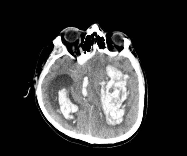
this sign may be confused with simultaneous intraparenchymal haemorrhages in multiple discrete locations which often occur within the deeper regions of the basal ganglia
when there are more than three satellite foci, this may be referred to as the island sign


 Unable to process the form. Check for errors and try again.