
Sublabral foramen
Citation, DOI, disclosures and article data
At the time the article was created Frank Gaillard had no recorded disclosures.
View Frank Gaillard's current disclosuresAt the time the article was last revised Lam Van Le had no financial relationships to ineligible companies to disclose.
View Lam Van Le's current disclosures- Sub-labral foramen
- Sublabral hole
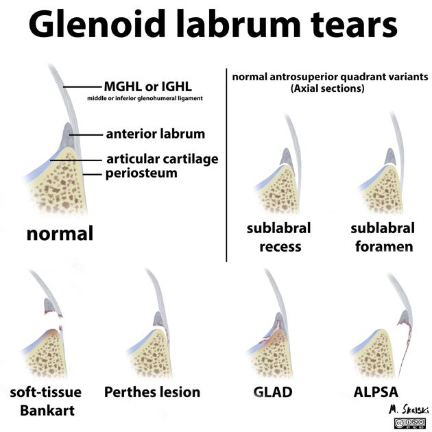
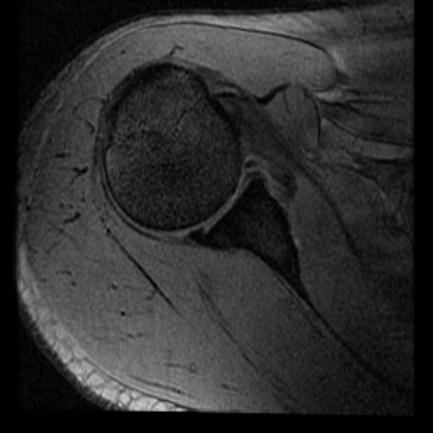
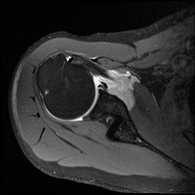
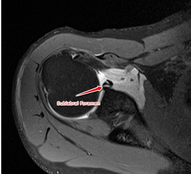
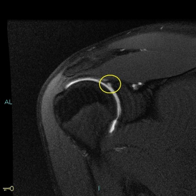
A sublabral foramen is simply a focal detachment of the anterosuperior labrum from the underlying glenoid and constitutes a normal labral variant of no clinical significance 1-4.
On imaging, it might be confused with a SLAP lesion or an anterior labral tear 1.
On this page:
Gross anatomy
Sublabral foramina are located anterosuperiorly and can extend down to but usually not below the level of the 3 o'clock position 1,3. It provides a communication between the glenohumeral joint and the subscapularis recess 1 and might be associated with a superior sublabral recess 1,4.
Radiographic features
MRI / MRA
On MRI a sublabral foramen can be seen as a focal detachment anterosuperiorly between the 1- o’clock to 3-o’clock position up to one or rarely two axial sections below the midpoint at the level of the anterior glenoid notch 3.
Clinical importance
Sublabral foramina are fairly frequent findings on MRI and might be found in up to 10-20% of normal patients 1,5,6. It is different from the superior sublabral sulcus or recess which can be found more superiorly underneath the long head biceps tendon origin 4. It might be also confused with a type II SLAP lesion or an anterior labral tear 3. Unlike those two the sublabral foramen is characterised by smoother contours, lacks significant displacement (>2 mm) and does not extend posteriorly past the insertion of the long head of the biceps tendon 3.
References
- 1. Dunham K, Bencardino J, Rokito A. Anatomic Variants and Pitfalls of the Labrum, Glenoid Cartilage, and Glenohumeral Ligaments. Magn Reson Imaging Clin N Am. 2012;20(2):213-28, x. doi:10.1016/j.mric.2012.01.014 - Pubmed
- 2. De Maeseneer M, Van Roy F, Lenchik L et al. CT and MR Arthrography of the Normal and Pathologic Anterosuperior Labrum and Labral-Bicipital Complex. Radiographics. 2000;20(suppl_1):S67-81. doi:10.1148/radiographics.20.suppl_1.g00oc03s67 - Pubmed
- 3. Tuite MJ, Blankenbaker DG, Seifert M et-al. Sublabral foramen and buford complex: inferior extent of the unattached or absent labrum in 50 patients. Radiology. 2002;223 (1): 137-42. doi:10.1148/radiol.2231010896 - Pubmed citation
- 4. Marcon G & Macedo T. Artifacts and Pitfalls in Shoulder Magnetic Resonance Imaging. Radiol Bras. 2015;48(4):242-8. doi:10.1590/0100-3984.2013.0006 - Pubmed
- 5. Rao A, Kim T, Chronopoulos E, McFarland E. Anatomical Variants in the Anterosuperior Aspect of the Glenoid Labrum: A Statistical Analysis of Seventy-Three Cases. J Bone Joint Surg Am. 2003;85(4):653-9. doi:10.2106/00004623-200304000-00011 - Pubmed
- 6. Ilahi O, Cosculluela P, Ho D. Classification of Anterosuperior Glenoid Labrum Variants and Their Association with Shoulder Pathology. Orthopedics. 2008;31(3):226. doi:10.3928/01477447-20080301-18 - Pubmed
Incoming Links
Related articles: Anatomy: Upper limb
-
skeleton of the upper limb
- clavicle
- scapula
- humerus
- radius
- ulna
- hand
- accessory ossicles of the upper limb
- accessory ossicles of the shoulder
- accessory ossicles of the elbow
-
accessory ossicles of the wrist (mnemonic)
- os centrale carpi
- os epilunate
- os epitriquetrum
- os styloideum
- os hamuli proprium
- lunula
- os triangulare
- trapezium secondarium
- os paratrapezium
- os radiostyloideum (persistent radial styloid)
- joints of the upper limb
-
pectoral girdle
-
shoulder joint
- articulations
- associated structures
- joint capsule
- bursae
- ligaments
- movements
- scapulothoracic joint
-
glenohumeral joint
- arm flexion
- arm extension
- arm abduction
- arm adduction
- arm internal rotation (medial rotation)
- arm external rotation (lateral rotation)
- circumduction
- arterial supply - scapular anastomosis
- ossification centres
-
shoulder joint
-
elbow joint
- proximal radioulnar joint
- ligaments
- associated structures
- movements
- alignment
- arterial supply - elbow anastomosis
- development
-
wrist joint
- articulations
-
ligaments
- intrinsic ligaments
- extrinsic ligaments
- radioscaphoid ligament
- dorsal intercarpal ligament
- dorsal radiotriquetral ligament
- dorsal radioulnar ligament
- volar radioulnar ligament
- radioscaphocapitate ligament
- long radiolunate ligament
- Vickers ligament
- short radiolunate ligament
- ulnolunate ligament
- ulnotriquetral ligament
- ulnocapitate ligament
- ulnar collateral ligament
- associated structures
- extensor retinaculum
- flexor retinaculum
- joint capsule
- movements
- alignment
- ossification centres
-
hand joints
- articulations
- carpometacarpal joint
-
metacarpophalangeal joints
- palmar ligament (plate)
- collateral ligament
-
interphalangeal joints
- palmar ligament (plate)
- collateral ligament
- movements
- ossification centres
- articulations
-
pectoral girdle
- spaces of the upper limb
- muscles of the upper limb
- shoulder girdle
- anterior compartment of the arm
- posterior compartment of the arm
-
anterior compartment of the forearm
- superficial
- intermediate
- deep
-
posterior compartment of the forearm (extensors)
- superficial
- deep
- muscles of the hand
-
accessory muscles
- elbow
- volar wrist midline
- palmaris longus profundus
- aberrant palmaris longus
- volar wrist radial-side
- accessory flexor digitorum superficialis indicis
- flexor indicis profundus
- flexor carpi radialis vel profundus
- accessory head of the flexor pollicis longus (Gantzer muscle, common)
- volar wrist ulnar-side
- dorsal wrist
- blood supply to the upper limb
-
arteries
- subclavian artery (mnemonic)
- axillary artery
- brachial artery (proximal portion)
- ulnar artery
- radial artery
- veins
-
arteries
- innervation of the upper limb
- intercostobrachial nerve
-
brachial plexus (mnemonic)
- branches from the roots
- branches from the trunks
- branches from the cords
- lateral cord
- posterior cord
- medial cord
- terminal branches
- lymphatic drainage of the upper limb


 Unable to process the form. Check for errors and try again.