
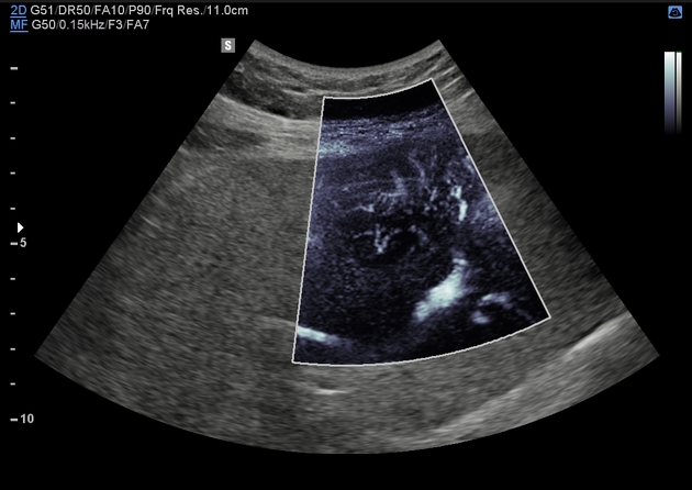
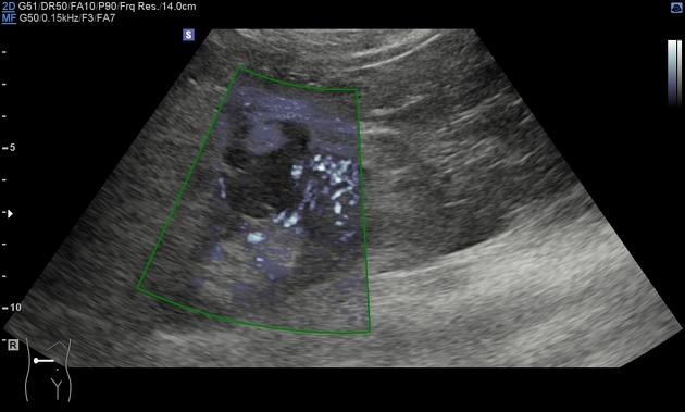
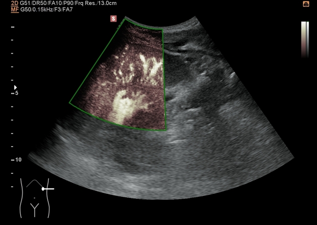
Superb microvascular imaging (SMI) or microvascular flow imaging (MVI/MV-flow - the name varying by manufacturers) is a ultrasound imaging technique that aims to visualise low velocity and small diameter blood vessel flow. Unlike conventional colour and power Doppler imaging, superb microvascular imaging can suppress noise caused by motion artifacts without removing the weak signal arising from small vessel blood flow, hence it achieves a greater sensitivity than those 1.
Superb microvascular imaging (similarly to power Doppler) typically displays a monochrome map of blood flow superimposed on the B-mode image, though some systems are now capable of providing colour-coded directional information as well.
Clinical use
Due to the relative novelty of the superb microvascular imaging (SMI) technique, strong clinical evidence about its use is lacking. Existing research shows that it can be utilised to:
characterise focal and diffuse liver lesions
assess vascularity of cystic and solid renal lesions 3,4
assess vascularity of breast masses 5
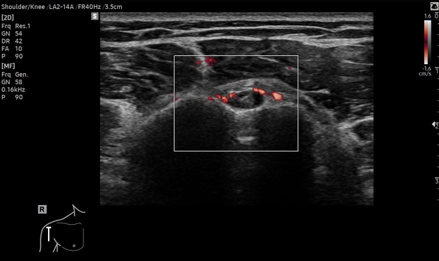
evaluate thyroid nodules
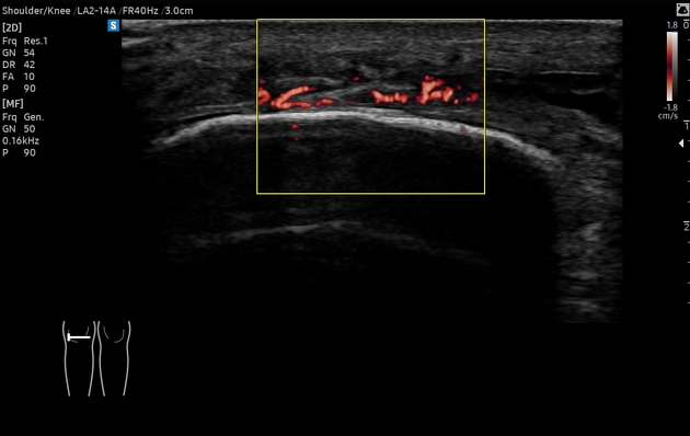
Superb microvascular imaging also shows promise as an adjunctive tool in musculoskeletal ultrasound by allowing more sensitive detection of increased vascularity in tendons, joint capsules, and peripheral nerves. Some studies have also demonstrated its potential use in evaluating intraplaque neovascularisation and thereby in assessing the risk of haemorrhage in carotid plaques 2.
Practical points
Several similar but proprietary microvascular flow imaging methods have been developed by different manufacturers, thus a similar technique can be encountered under various names in the literature:
microvascular flow (MV-flow, Samsung Medison)
microvascular imaging (MVI, General Electric)
superb microvascular imaging (SMI, Canon Medical Systems)
-
Slow flow (Siemens Healthineers)


 Unable to process the form. Check for errors and try again.