
Thrombolysis in cerebral infarction (TICI) scale
Citation, DOI, disclosures and article data
At the time the article was created Craig Hacking had no recorded disclosures.
View Craig Hacking's current disclosuresAt the time the article was last revised Jeremy Jones had no financial relationships to ineligible companies to disclose.
View Jeremy Jones's current disclosures- TICI
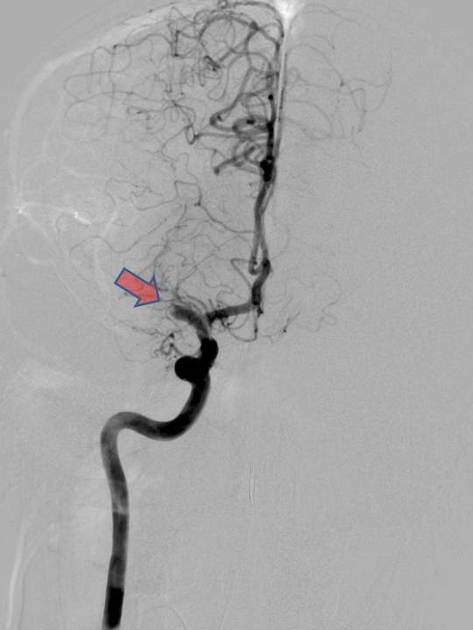
The thrombolysis in cerebral infarction (TICI) grading system was described in 2003 by Higashida et al. 1 as a tool for determining the response of thrombolytic therapy for ischaemic stroke. In neurointerventional radiology it is commonly used for patients post endovascular revascularisation. Like most therapy response grading systems, it predicts prognosis.
Classification
The original description 1 was based on the angiographic appearances of the treated occluded vessel and the distal branches:
grade 0: no perfusion
grade 1: penetration with minimal perfusion
-
grade 2: partial perfusion
grade 2A: only partial filling (less than two-thirds) of the entire vascular territory is visualised
grade 2B: complete filling of all of the expected vascular territory is visualised but the filling is slower than normal
grade 3: complete perfusion
In 2013 Fugate et al. reported marked variability in its definitions and application 2. Furthermore, a consensus paper from three collaborative groups published in Stroke in 2013 3 recommended a modified scale, and a change of name to modified treatment in cerebral infarction (mTICI), to better reflect the increased use of endovascular therapies.
See also
References
- 1. Higashida R, Furlan A, Roberts H et al. Trial Design and Reporting Standards for Intra-Arterial Cerebral Thrombolysis for Acute Ischemic Stroke. Stroke. 2003;34(8):e109-37. doi:10.1161/01.STR.0000082721.62796.09 - Pubmed
- 2. Fugate J, Klunder A, Kallmes D. What is Meant by "TICI"? AJNR Am J Neuroradiol. 2013;34(9):1792-7. doi:10.3174/ajnr.A3496 - Pubmed
- 3. Zaidat O, Yoo A, Khatri P et al. Recommendations on Angiographic Revascularization Grading Standards for Acute Ischemic Stroke: A Consensus Statement. Stroke. 2013;44(9):2650-63. doi:10.1161/STROKEAHA.113.001972 - Pubmed
Incoming Links
- Vertebrobasilar artery occlusion - diagnosis and treatment
- Bilateral ACA infarction due to azygos ACA embolism
- Acute P1 occlusion with PCA ischaemic penumbra (CT perfusion)
- Acute M1 occlusion with ischaemic penumbra (CT perfusion)
- Proximal right MCA M1 segment embolic occlusion
- Acute left middle cerebral artery territory infarct with clot retrieval
- Left MCA stroke for ECR - tortuous access
- Left MCA stroke with tandem carotid occlusion
- Right MCA territory infarction
Related articles: Stroke and intracranial haemorrhage
-
stroke and intracranial haemorrhage
- general articles
-
ischaemic stroke
- general discussions
- scoring and classification systems
- Alberta stroke program early CT score (ASPECTS)
- ASCOD classification
- Canadian Neurological Scale
- Heidelberg bleeding classification
- NIH Stroke Scale
- Mathew stroke scale
- modified Rankin scale
- Orgogozo Stroke Scale
- Scandinavian Stroke Scale
- thrombolysis in cerebral infarction (TICI) scale
- TOAST classification
- collateral vessel scores
- signs
- by region
- hemispheric infarcts
- frontal lobe infarct
- parietal lobe infarct
- temporal lobe infarct
- occipital lobe infarct
- alexia without agraphia syndrome: PCA
- cortical blindness syndrome (Anton syndrome): top of basilar or bilateral PCA
- Balint syndrome: bilateral PCA
- lacunar infarct
-
thalamic infarct
- artery of Percheron infarct
- Déjerine-Roussy syndrome (thalamic pain syndrome): thalamoperforators of PCA
- top of the basilar syndrome
- striatocapsular infarct
- choroid plexus infarct
- cerebellar infarct
-
brainstem infarct
- midbrain infarct
- Benedikt syndrome: PCA
- Claude syndrome: PCA
- Nothnagel syndrome: PCA
- Weber syndrome: PCA
- Wernekink commissure syndrome
- pontine infarct
- Brissaud-Sicard syndrome
- facial colliculus syndrome
- Gasperini syndrome: basilar artery or AICA
- inferior medial pontine syndrome (Foville syndrome): basilar artery
- lateral pontine syndrome (Marie-Foix syndrome): basilar artery or AICA
- locked-in syndrome: basilar artery
- Millard-Gubler syndrome: basilar artery
- Raymond syndrome: basilar artery
- medullary infarct
- Babinski-Nageotte syndrome
- Cestan-Chenais syndrome
- hemimedullary syndrome (Reinhold syndrome)
- lateral medullary stroke syndrome (Wallenberg syndrome)
- medial medullary syndrome (Déjerine syndrome)
- Opalski syndrome
- midbrain infarct
- acute spinal cord ischaemia syndrome
- hemispheric infarcts
- by vascular territory
- by vessel size
- treatment options
- complications
-
intracranial haemorrhage
-
intra-axial haemorrhage
- signs and formulas
- ABC/2 (volume estimation)
- black hole sign
- blend sign
- cashew nut sign
- CTA spot sign
- island sign
- satellite sign
- swirl sign
- zebra sign
- by type
- by location
- signs and formulas
- extra-axial haemorrhage
- extradural haemorrhage (EDH)
- intralaminar dural haemorrhage
- subdural haemorrhage (SDH)
-
subarachnoid haemorrhage (SAH)
- types
- complications
- grading systems
- subpial haemorrhage
-
intra-axial haemorrhage


 Unable to process the form. Check for errors and try again.