
Tracheal diverticulum
Citation, DOI, disclosures and article data
At the time the article was created Laughlin Dawes had no recorded disclosures.
View Laughlin Dawes's current disclosuresAt the time the article was last revised Tariq Walizai had no financial relationships to ineligible companies to disclose.
View Tariq Walizai's current disclosures- Tracheal diverticula
- Diverticulum of trachea
- Tracheocele
- Tracheoceles
- Tracheocoele
- Tracheocoeles
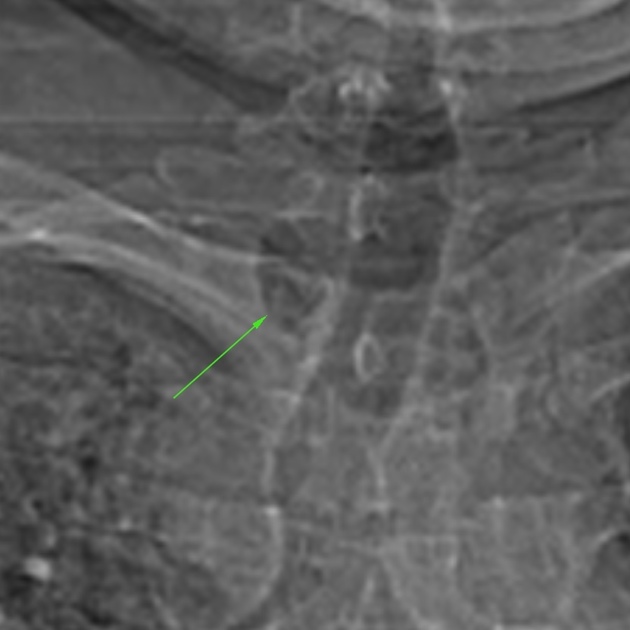
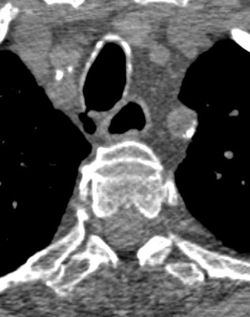
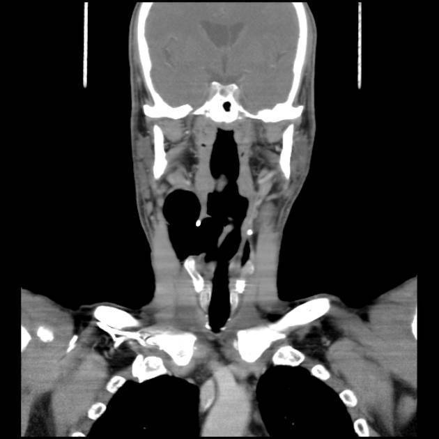
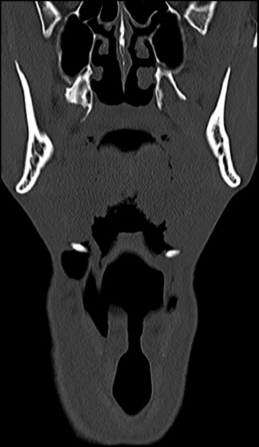
Tracheal diverticula, also known as tracheoceles, are usually an incidental finding. Occasionally they may mimic pneumomediastinum, a so-called pseudopneumomediastinum.
On this page:
Terminology
There is an overlap in the use and description of the terms paratracheal air cyst and a tracheal diverticulum in the literature such that they often seem to be synonyms for the same entity, which they are not 1,5.
Epidemiology
Associations
chronic obstructive pulmonary disease (COPD): for acquired type 3
tracheobronchomegaly: when multiple tracheal diverticula are present 5
Clinical presentation
Tracheal diverticula are typically asymptomatic but can occasionally present with chronic cough, stridor, dyspnoea, or recurrent infection 5.
Pathology
Tracheal diverticula can be 5:
congenital
-
acquired
prolonged increased intraluminal pressure, e.g. chronic cough, COPD
iatrogenic, e.g. post-surgical
Location
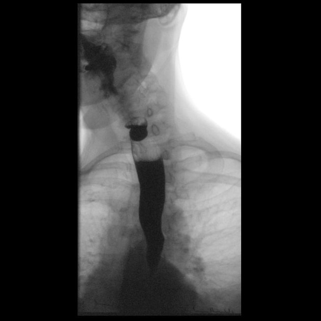
A tracheal diverticulum projects posteriorly where the cartilage rings of the trachea are deficient and usually lies to the right where there is no oesophagus supporting the paratracheal tissue 6. The vast majority (97%) are located at the right posterolateral aspect of the trachea, approximately at the level of the thoracic inlet 5. A direct connection with the trachea may not always be appreciable on CT 5.
ADVERTISEMENT: Supporters see fewer/no ads
Treatment and prognosis
Although usually asymptomatic, tracheal diverticula may accumulate respiratory secretions that become infected (and potentially abscess-forming) and lead to coughing or tracheobronchitis.
History and etymology
In 1838, Rokitansky described three cases of tracheal diverticula for the first time 7.
Differential diagnosis
Quiz questions
References
- 1. Buterbaugh J & Erly W. Paratracheal Air Cysts: A Common Finding on Routine CT Examinations of the Cervical Spine and Neck That May Mimic Pneumomediastinum in Patients with Traumatic Injuries. AJNR Am J Neuroradiol. 2008;29(6):1218-21. doi:10.3174/ajnr.A1058 - Pubmed
- 2. Goo J, Im J, Ahn J et al. Right Paratracheal Air Cysts in the Thoracic Inlet: Clinical and Radiologic Significance. AJR Am J Roentgenol. 1999;173(1):65-70. doi:10.2214/ajr.173.1.10397101 - Pubmed
- 3. Webb E, Elicker B, Webb W. Using CT to Diagnose Nonneoplastic Tracheal Abnormalities: Appearance of the Tracheal Wall. AJR Am J Roentgenol. 2000;174(5):1315-21. doi:10.2214/ajr.174.5.1741315 - Pubmed
- 4. Higuchi T, Takahashi N, Shiotani M, Maeda H, Yoshimura N. Characteristics and CT Features of Subcarinal Air Collections/main Bronchial Diverticula. Eur Radiol. 2010;20(1):95-9. doi:10.1007/s00330-009-1526-z - Pubmed
- 5. Tanrivermis Sayit A, Elmali M, Saglam D, Celenk C. The Diseases of Airway-Tracheal Diverticulum: A Review of the Literature. J Thorac Dis. 2016;8(10):E1163-7. doi:10.21037/jtd.2016.10.92 - Pubmed
- 6. Pace M, Dapoto A, Surace A et al. Tracheal Diverticula: A Retrospective Analysis of Patients Referred for Thoracic CT. Medicine (Baltimore). 2018;97(39):e12544. doi:10.1097/MD.0000000000012544 - Pubmed
- 7. Mathey J & Lemoine A. Tracheal Diverticulum and Congenital Oesophagotracheal Fistula Without Oesophageal Atresia. Thorax. 1954;9(2):106-11. doi:10.1136/thx.9.2.106 - Pubmed
Incoming Links
- Laryngeal amyloidosis
- Hydropneumothorax
- Tracheal diverticulum
- Tracheal diverticulum
- Tracheal diverticulum
- Accidental foreign body aspiration (seamstress needle)
- Gemcitabine lung toxicity
- Tracheal diverticulum
- Tracheal diverticulum
- Birt-Hogg-Dubé syndrome
- Tracheal web - low-dose lung cancer screening (CT)
- Tracheal diverticulum
- Paratracheal air cyst
- Mounier-Kuhn syndrome
- Tracheal diverticulum
Related articles: Chest
- imaging techniques
-
chest radiograph
- radiography
-
approach
- ABCDE
- ABCDEFGHI
- congenital heart disease
- medical devices in the thorax
- common lines and tubes
- nasogastric tubes
- endotracheal tubes
- central venous catheters
- oesophageal temperature probe
- tracheostomy tube
- pleural catheters
- cardiac conduction devices
- prosthetic heart valve
- review areas
-
airspace opacification
- differential diagnoses of airspace opacification
- lobar consolidation
-
atelectasis
- mechanism-based
- morphology-based
- lobar lung collapse
- chest x-ray in the exam setting
- cardiomediastinal contour
- chest radiograph zones
- tracheal air column
- fissures
- normal chest x-ray appearance of the diaphragm
- nipple shadow
-
lines and stripes
- anterior junction line
- posterior junction line
- right paratracheal stripe
- left paratracheal stripe
- posterior tracheal stripe/tracheo-oesophageal stripe
- posterior wall of bronchus intermedius
- right paraspinal line
- left paraspinal line
- aortic-pulmonary stripe
- aortopulmonary window
- azygo-oesophageal recess
- spaces
- signs
- air bronchogram
- big rib sign
- Chang sign
- Chen sign
- coin lesion
- continuous diaphragm sign
- dense hilum sign
- double contour sign
- egg-on-a-string sign
- extrapleural sign
- finger in glove sign
- flat waist sign
- Fleischner sign
- ginkgo leaf sign
- Golden S sign
- Hampton hump
- haystack sign
- hilum convergence sign
- hilum overlay sign
- Hoffman-Rigler sign
- holly leaf sign
- incomplete border sign
- juxtaphrenic peak sign
- Kirklin sign
- medial stripe sign
- melting ice cube sign
- more black sign
- Naclerio V sign
- Palla sign
- pericardial fat tag sign
- Shmoo sign
- silhouette sign
- snowman sign
- spinnaker sign
- steeple sign
- straight left heart border sign
- third mogul sign
- tram-track sign
- walking man sign
- water bottle sign
- wave sign
- Westermark sign
- HRCT
-
chest radiograph
- airways
- bronchitis
- small airways disease
-
bronchiectasis
- broncho-arterial ratio
- related conditions
- differentials by distribution
- narrowing
-
tracheal stenosis
- diffuse tracheal narrowing (differential)
-
bronchial stenosis
- diffuse airway narrowing (differential)
-
tracheal stenosis
- diverticula
- pulmonary oedema
-
interstitial lung disease (ILD)
- Anti-Jo-1 antibody-positive interstitial lung disease
- drug-induced interstitial lung disease
-
hypersensitivity pneumonitis
- acute hypersensitivity pneumonitis
- subacute hypersensitivity pneumonitis
- chronic hypersensitivity pneumonitis
- aetiology
- bird fancier's lung: pigeon fancier's lung
- farmer's lung
- cheese workers' lung
- bagassosis
- mushroom worker’s lung
- malt worker’s lung
- maple bark disease
- hot tub lung
- wine maker’s lung
- woodsman’s disease
- thatched roof lung
- tobacco grower’s lung
- potato riddler’s lung
- summer-type pneumonitis
- dry rot lung
- machine operator’s lung
- humidifier lung
- shower curtain disease
- furrier’s lung
- miller’s lung
- lycoperdonosis
- saxophone lung
-
idiopathic interstitial pneumonia (mnemonic)
- acute interstitial pneumonia (AIP)
- cryptogenic organising pneumonia (COP)
- desquamative interstitial pneumonia (DIP)
- non-specific interstitial pneumonia (NSIP)
- idiopathic pleuroparenchymal fibroelastosis
- lymphoid interstitial pneumonia (LIP)
- respiratory bronchiolitis–associated interstitial lung disease (RB-ILD)
- usual interstitial pneumonia / idiopathic pulmonary fibrosis (UIP/IPF)
-
pneumoconioses
- fibrotic
- non-fibrotic
-
lung cancer
-
non-small-cell lung cancer
-
adenocarcinoma
- pre-invasive tumours
- minimally invasive tumours
- invasive tumours
- variants of invasive carcinoma
- described imaging features
- adenosquamous carcinoma
- large cell carcinoma
- primary sarcomatoid carcinoma of the lung
- squamous cell carcinoma
- salivary gland-type tumours
-
adenocarcinoma
- pulmonary neuroendocrine tumours
- preinvasive lesions
-
lung cancer invasion patterns
- tumour spread through air spaces (STAS)
- presence of non-lepidic patterns such as acinar, papillary, solid, or micropapillary
- myofibroblastic stroma associated with invasive tumour cells
- pleural invasion
- vascular invasion
- tumours by location
- benign neoplasms
- pulmonary metastases
- lung cancer screening
- lung cancer staging
-
non-small-cell lung cancer


 Unable to process the form. Check for errors and try again.