
A triceps tendon rupture represents the extreme end of the spectrum of triceps tendon tears where there is complete detachment of the triceps tendon. It most often occurs at the distal end. It is more common in males and seen in age groups 30-50.
Pathology
If can either occur in an acute setting with trauma (e.g. as a result of a sudden forceful elbow contraction in weightlifters) or on a background of chronic triceps tendinopathic changes (older individuals with underlying systemic illnesses).
A rupture most commonly occurs at the osseous insertion of the medial or lateral head as a detachment of tendon from the bone.
Musculotendinous junction or intramuscular ruptures are considered very rare 4.
Risk factors
Chronic conditions which include
chronic renal insufficiency requiring dialysis
metabolic bone diseases leading to hyperparathyroidism
chronic olecranon bursitis: can increases the risk of rupture due to chronic inflammation around the tendon
underlying elbow arthroplasty
Radiographic features
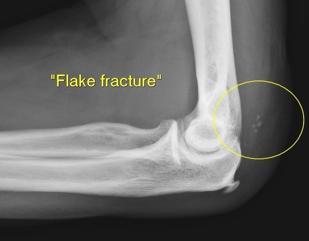
Plain film
Non-specific but may show an appearance of a "flake sign" or "fleck sign" on a lateral radiograph.
Ultrasound
May show a gap in tendon with lack of movement on dynamic interrogation +/- fusiform swelling and retraction of tendon / muscle portions.
MRI
MRI may overestimate tears 1 but may show a deficit - gap in the tendon +/- surrounding soft tissue inflammatory changes. MRI can be beneficial in differentiating between partial and complete tears.


 Unable to process the form. Check for errors and try again.