
Presentation
Weight lifter with pain and swelling at the back of the left elbow after a fall on an outstretched hand.
Patient Data
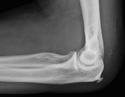
Lateral view showing the “flake” fracture avulsed from the olecranon. There is also enthesopathy at the triceps insertion.
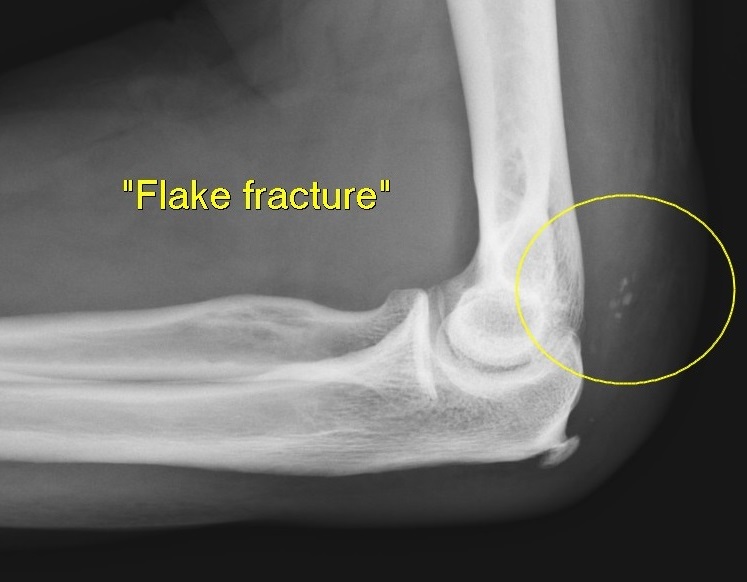
Lateral view showing the “flake” fracture avulsed from the olecranon (yellow circle)
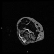

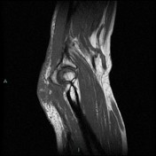
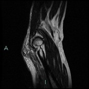
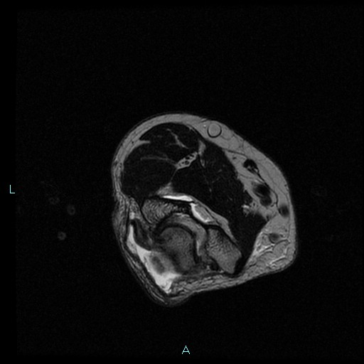
MRI left elbow
The posterior component of the distal triceps tendon (combined tendon of the lateral and long heads) is completely avulsed and retracted. The anterior or deep component of the distal triceps (medial head) remains intact. The posterior or superficial component is retracted of 3 cm and the deep one is continuous and intact with fluid filled gap where the triceps tendon is devoid and retracted.
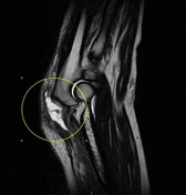
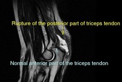

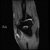
The posterior component of the distal triceps tendon (combined tendon of the lateral and long heads) is completely avulsed and retracted. The anterior or deep component of the distal triceps (medial head) remains intact. The posterior or superficial component is retracted of 3 cm and the deep one is continuous and intact with fluid filled gap where the triceps tendon is devoid and retracted (yellow circle).
Case Discussion
The triceps is a tripennate muscle with origins on the infraglenoid tubercle of the scapula (long head), the upper posterior humerus (lateral head), and lower posterior humerus (medial head). The distal insertion is primarily on the olecranon, with a lateral aponeurotic expansion that extends distally over the anconeus to the proximal forearm fascia. Rupture of the triceps tendon usually occurs at or near the insertion of the tendon onto the olecranon, as a result of an active contraction of the triceps in extension with a forced passive flexion. Rupture is often associated with pre-existing systemic conditions or drug treatments, including the local or systemic of steroids, anabolic steroid use, local steroid injection. Treatment depends on the extent of the rupture, level of activity and age of the patient. Good results of conservative treatment is often effective in these cases. X ray on lateral view may show small fleck of bone avulsed from the olecranon "flake sign".


 Unable to process the form. Check for errors and try again.