
Urachus
Citation, DOI, disclosures and article data
At the time the article was created Ian Bickle had no recorded disclosures.
View Ian Bickle's current disclosuresAt the time the article was last revised Craig Hacking had the following disclosures:
- Philips Australia, Paid speaker at Philips Spectral CT events (ongoing)
These were assessed during peer review and were determined to not be relevant to the changes that were made.
View Craig Hacking's current disclosures- Median umbilical ligaments
- Median umbilical ligament
- Urachuses
- Urachi
The urachus (plural: urachuses or urachi 6,7) is the fibrous vestigial remnant of the embryonic allantois.
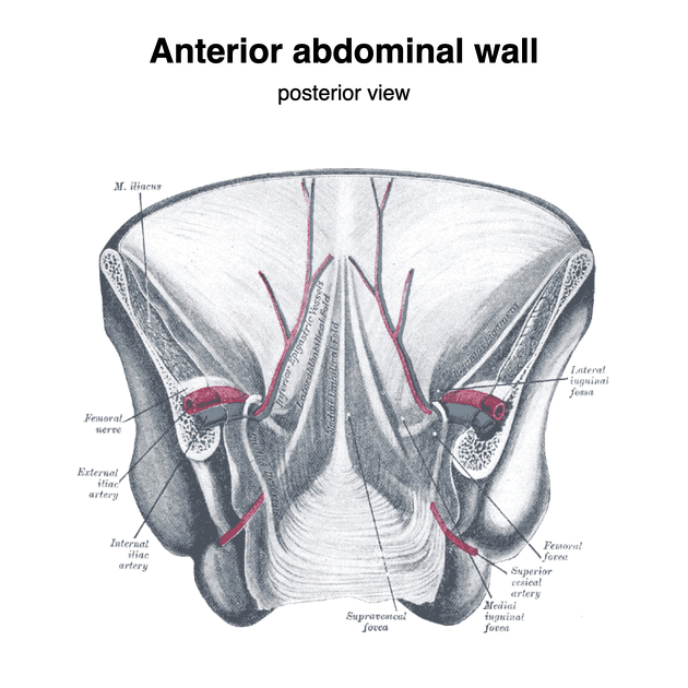
The lumen of the urachus usually obliterates following birth and becomes known as the median umbilical ligament which is in turn covered by a midline linear fibrous fold of parietal peritoneum (the median umbilical fold) 3. This fold extends from the apex of the bladder to the umbilicus. It is located in the retropubic space.
If the lumen does not completely involute, a spectrum of urachal remnants may persist, including 4:
A urachal remnant may transform into an adenocarcinoma 5.
References
- 1. Yu JS, Kim KW, Lee HJ, Lee YJ, Yoon CS, Kim MJ. Urachal remnant diseases: spectrum of CT and US findings. (2001) Radiographics : a review publication of the Radiological Society of North America, Inc. 21 (2): 451-61. doi:10.1148/radiographics.21.2.g01mr02451 - Pubmed
- 2. Moore KL, Agur AMR, Dalley AF. Clinically oriented anatomy. LWW. ISBN:1451119453. Read it at Google Books - Find it at Amazon
- 3. Last's anatomy, regional and applied. Churchill Livingstone. ISBN:044304662X. Read it at Google Books - Find it at Amazon
- 4. Parada Villavicencio C, Adam SZ, Nikolaidis P, Yaghmai V, Miller FH. Imaging of the Urachus: Anomalies, Complications, and Mimics. Radiographics : a review publication of the Radiological Society of North America, Inc. 36 (7): 2049-2063. doi:10.1148/rg.2016160062 - Pubmed
- 5. Jandou, I., Ettanji, A., Mohammed, E., Moataz, A., Mohammed, D., Debbagh, A., & Aboutaieb, R. (2021). Mucosal urinary secretion revealing adenocarcinoma of urachus. Annals of Medicine and Surgery, 65, 102335. doi:10.1016/j.amsu.2021.102335
- 6. Liang Cheng, David G. Bostwick. Urologic Surgical Pathology E-Book. (2014) ISBN: 9780323086196
- 7. Yiee, Jenny & Garcia, Nilda & Baker, Linda & Barber, Robert & Snodgrass, Warren & Wilcox, Duncan. (2008). A diagnostic algorithm for urachal anomalies. Journal of pediatric urology. 3. 500-4. 10.1016/j.jpurol.2007.07.010. doi:10.1016/j.jpurol.2007.07.010
Incoming Links
- Infected urachal cyst with sinus formation
- Urachal adenocarcinoma
- Prominent median umbilical ligament
- Median umbilical ligament
- Renal cell carcinoma
- Patent urachus - adult
- Intravesical urachal cyst
- Urachal adenocarcinoma
- Urachus (illustration)
- Vesicourachal diverticulum
- Infected urachal cyst
- Infected patent urachus
- Median umbilical ligament
- Urachal remnant adenocarcinoma
Related articles: Pathology: Genitourinary
- obstetrics
-
first trimester
- ultrasound findings in early pregnancy
- embryo/fetus
- beta-hCG levels
- confirming intrauterine gestation
- pregnancy of unknown location (PUL)
- first trimester vaginal bleeding
- early structural scan
- aneuploidy testing
-
second trimester
- fetal biometry
- amniotic fluid volume
- fetal morphology assessment
- soft markers
- amnioreduction
- Doppler ultrasound
- nuchal translucency
- 11-13 weeks antenatal scan
- chorionic villus sampling (CVS) and amniocentesis
- other
- placenta
- placental anatomy
- placental developmental abnormalities
- placenta praevia
- spectrum of abnormal placental villous adherence
- abnormalities of cord insertion
- abruptio placentae
- placental pathology
- vascular pathologies of placenta
- placental infections
- placental masses
- molar pregnancy
- twin placenta
- miscellaneous
-
first trimester
- gynaecology
- acute pelvic pain
- chronic pelvic pain
- uterus
- ovaries
- ovarian follicle
- ovarian torsion
- pelvic inflammatory disease
- ovarian cysts and masses
- paraovarian cyst
- polycystic ovaries
- ovarian hyperstimulation syndrome
- post-hysterectomy ovary
- cervix
- fallopian tube
- other
- male genital tract
- prostate gland
- transrectal ultrasound
- prostate tumours
- infections of the prostate
-
prostatitis
- acute bacterial prostatitis
-
chronic prostatitis
- chronic bacterial prostatitis
- chronic prostatitis and chronic pelvic pain syndrome (CPPS)
- asymptomatic inflammatory prostatitis
- granulomatous prostatitis
- emphysematous prostatitis
- prostatic abscess
-
prostatitis
- benign prostatic hypertrophy
- cystic lesions of the prostate
- prostatic calcification
- prostatic infarction
- testes
-
unilateral testicular lesion
- testicular torsion
- orchitis
- testicular trauma
-
germ cell tumours of the testis
- testicular seminoma
-
non seminomatous germ cell tumours
- mixed germ cell tumour
- yolk sac tumour (endodermal sinus tumour)
- embryonal cell carcinoma
- choriocarcinoma
- testicular teratoma
- testicular epidermoid (teratoma with ectodermal elements only)
- burned out testis tumour
- sex cord / stromal tumours of the testis
- testicular cyst
- testicular lymphoma
- bilateral testicular lesion
- paratesticular lesions
- epididymis
- other
- polyorchidism
- cryptorchidism
- tubular ectasia of the rete testis
- cystadenoma of the rete testis
- testicular sarcoidosis
- testicular tuberculosis
- spermatic cord
- fibrous pseudotumour of the scrotum
- scrotal leiomyosarcoma
- testicular adrenal rest tumours (TARTs)
- tunica vaginalis testis mesothelioma
- splenogonadal fusion
- testicular vasculitis
- abnormal testicular Doppler flow (differential)
-
unilateral testicular lesion
- penis
- prostate gland
- KUB
- kidneys
- normal renal anatomy
- hydronephrosis
- urolithiasis
- renal masses
- renal cystic disease
- renal infection
- vascular
- trauma
- ureter
- normal ureter anatomy
- ureteral stricture
- ureteral dilatation
- ureteral anomalies
- ureteral tumours
- ureteral trauma
- other
- bladder
- kidneys


 Unable to process the form. Check for errors and try again.