
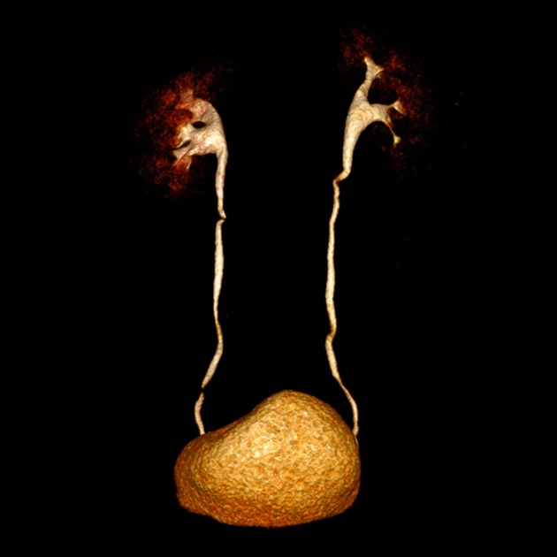
The ureter is a paired fibromuscular tube that conveys urine from the kidneys in the abdomen to the bladder in the pelvis.
On this page:
Gross anatomy
The ureter is 25-30 cm long and has three parts:
abdominal ureter: from the renal pelvis to the pelvic brim
pelvic ureter: from the pelvic brim to the bladder
intravesical or intramural ureter: within the bladder wall
Course
The ureter begins its descent to the bladder by running along the medial aspect of the psoas muscle. Here, the ureter lies anteriorly and slightly medial to the tips of the L2-L5 transverse processes.
It enters the pelvis anteriorly to the sacroiliac joint at the bifurcation of the common iliac vessels (at the pelvic brim) and then courses anteriorly to the internal iliac artery down the lateral pelvic sidewall.
At the level of the ischial spine it turns forward and medially to enter the posterolateral wall of the bladder, where it runs an oblique 1-2 cm course, before opening into the bladder at the internal ureteric orifice 1,2.
Relations
The relations of the ureter are somewhat complex due to the differences between the left and right sides of the abdominal cavity and differences between male and female pelvic viscera.
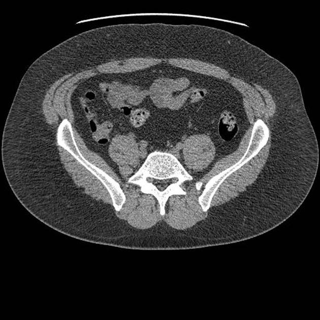
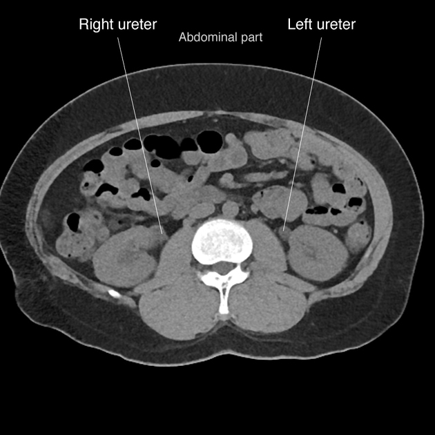
Abdominal ureter
Following the course of the ureter from superior to inferior 1,2:
posteriorly: psoas muscle; genitofemoral nerve; common iliac vessels; tips of L2-L5 transverse processes
-
anteriorly
right ureter: descending duodenum (D2); gonadal vessels; right colic vessels; ileocolic vessels, root of small bowel mesentery
left ureter: gonadal artery; left colic artery; loops of jejunum; apex of sigmoid mesocolon and colon
-
medially
right ureter: IVC
left ureter: abdominal aorta, inferior mesenteric vein
Pelvic ureter
posteriorly: sacroiliac joint, internal iliac artery
-
inferiorly 4
male: seminal vesicle
female: lateral fornix of the vagina
-
anteriorly 4
male: ductus deferens
female: uterine artery (in the broad ligament)
-
medially
female: cervix
It should be noted that the only structures to pass anteriorly over the pelvic ureter are the ductus deferens in males and the uterine artery in females.
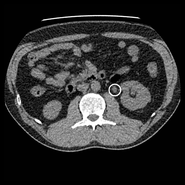
Constrictions
The ureter has a diameter of 3 mm 1 but there are three constrictions, which are the most common sites of renal calculus obstruction:
at the pelviureteric junction (PUJ) of the renal pelvis and the ureter
as the ureter enters the pelvis and crosses over the common iliac artery bifurcation
at the vesicoureteric junction (VUJ) as the ureter obliquely enters the bladder wall
Blood supply
-
arterial supply: from branches of the renal artery, abdominal aorta, superior and inferior vesical arteries
some texts also include supply from the gonadal, middle rectal and uterine arteries 1,2
venous drainage: via similarly named veins but is highly variable 1,2
Lymphatic drainage
abdominal ureter: aorto-caval and common iliac nodes
pelvic ureter: internal and external iliac nodes 1
Innervation
autonomic innervation is derived from renal, aorticorenal and superior hypogastric plexuses 1
Histology
The ureteric wall is composed of three layers (from outside to inside):
adventitia
smooth muscle
transitional cell epithelium 1


 Unable to process the form. Check for errors and try again.