
Vagal paragangliomas (previously known as glomus vagale tumours) are tumours that occur along the path of the vagus nerve (CN X). They are a subset of extra-adrenal neuroendocrine tumours that are derived from the nonchromaffin paraganglion cells.
On this page:
Terminology
The term "glomus" was historically used to describe certain types of neuroendocrine tumours arising from paraganglia. The term is, however, imprecise and can be confused with the glomus bodies and tumours that arise from them. It can also be mixed up with glomus tumours of the subcutaneous skin, also referred to as glomangioma.
Clinical presentation
Typically presents as a painless mass behind the carotid artery. Vocal cord paralysis is a relatively frequent finding (~47%) 3.
Pathology
For a general discussion on the pathology of these tumours, please refer to the parental article on paragangliomas.
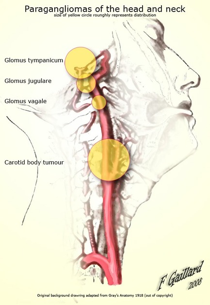
Location
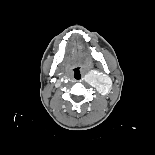
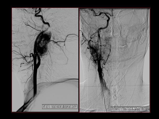
Although they could occur at a similar position to carotid body tumours they tend to be more rostral in location and do not widen the carotid bifurcation. They displace the internal and external carotid arteries anteriorly, and the internal jugular vein posteriorly 1.
Radiographic features
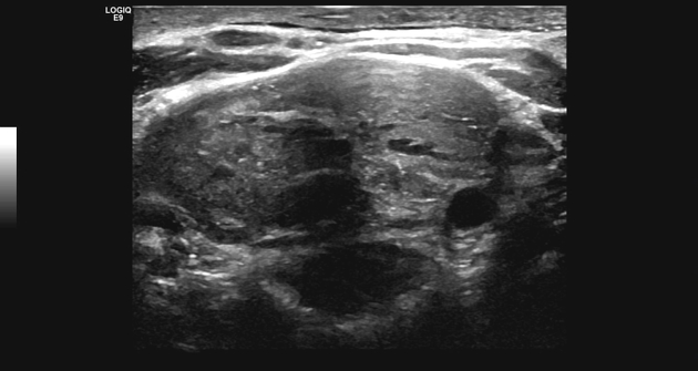
Ultrasound
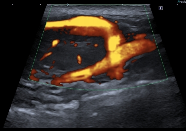
Difficult to sonographically differentiate between other lesions that can potentially occur in this location. It may be seen as a solid heterogeneously hypoechoic lesion comprising small vascular structures 3.
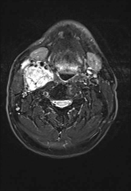
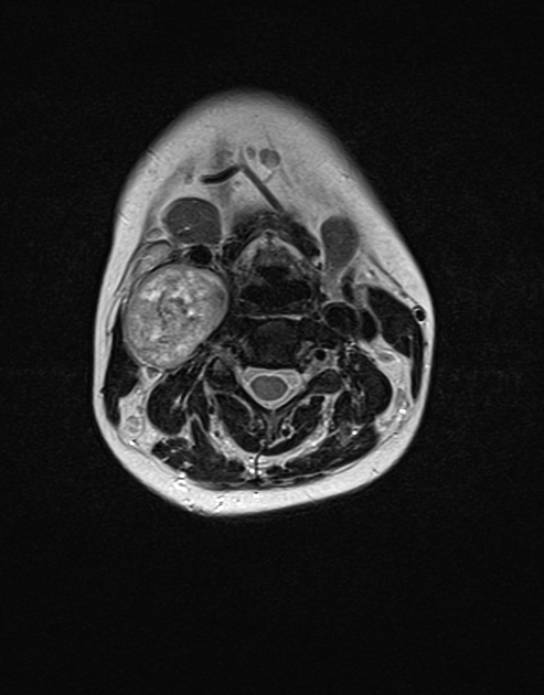
MRI
T1: usually low signal
T2: high signal with multiple flow voids, which may give a salt and pepper appearance
T1 C+ (Gd): intense enhancement
Differential diagnoses
The differential for lesions in this location include 2,4:
carotid space meningioma (extends from jugular foramen)


 Unable to process the form. Check for errors and try again.