
Presentation
Painless left neck swelling.
Patient Data
Age: 25 years
Gender: Male
From the case:
Carotid body tumor

Download
Info
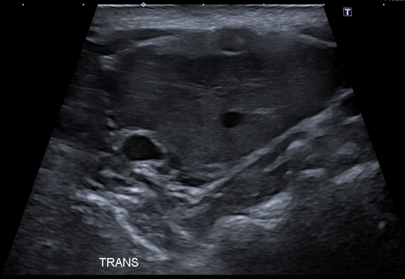
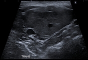
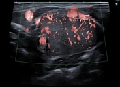
There is a well-defined homogeneous hypoechoic ovoid mass measuring (4.3 X 4.1 X 2.4 cm), splaying of the ICA and ECA. The color and power Doppler show prominent internal vascularity.
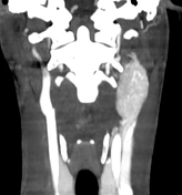
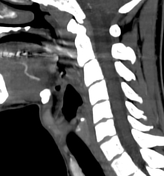
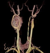
From the case:
Carotid body tumor

Download
Info
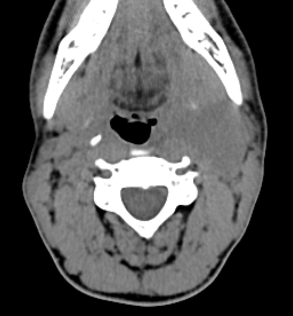
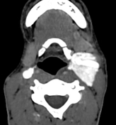
Left cervical soft tissue mass isodense to the muscles with strong enhancement, splaying the internal and external carotid arteries.
Case Discussion
The ultrasound and CT features are suggestive of a carotid body tumor.
Additional contributor: ZE. Boudiaf, MD, CHU Constantine


 Unable to process the form. Check for errors and try again.