
Visceral space
Citation, DOI, disclosures and article data
At the time the article was created Henry Knipe had no recorded disclosures.
View Henry Knipe's current disclosuresAt the time the article was last revised Kajanan Nithiyananthan had no financial relationships to ineligible companies to disclose.
View Kajanan Nithiyananthan's current disclosures- Visceral compartment
- Pretracheal space
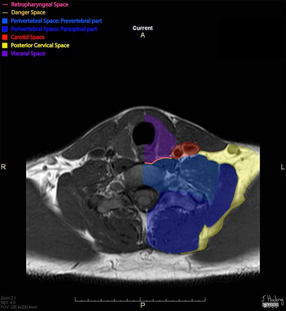
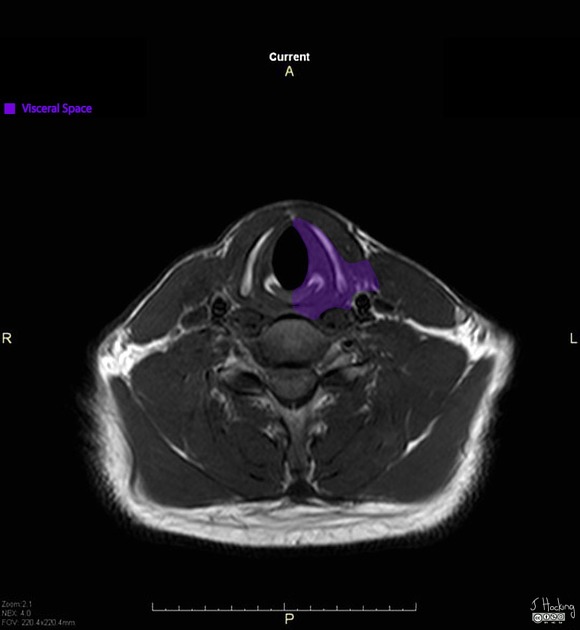
The visceral space or compartment is a deep compartment of the head and neck that contains the thyroid gland, larynx, trachea, upper oesophagus, hypopharynx and, in some definitions, oropharynx and nasopharynx.
On this page:
Images:
Terminology
Of the deep head and neck compartments, the visceral space has the most controversial terminology.
The main difference is the superior limit of the visceral space. Authors differ as to whether the visceral fascia, part of the middle layer of the deep cervical fascia, extends superiorly only to the hyoid bone or to the skull base 1. As such, some authors restrict the visceral space to the infrahyoid neck 2, including in this article, while other authorities subsume the entire pharyngeal mucosal space, including nasopharynx and oropharynx, as a subcomponent or synonym of the visceral space 1,3,4.
A second difference is the posterior limit of the visceral space. Some authorities divide the visceral space into an anterior "pretracheal space" and a posterior "retrovisceral space" 3, the latter of which is synonymous with the retropharyngeal space superiorly and retrooesophageal space inferiorly. In this definition, the entire visceral space is posteriorly delimited by the alar fascia, which is part of the deep layer of the deep cervical fascia, rather than by the visceral fascia. The rationale is that there is a free communication between these two spaces at the level between the thyroid cartilage and the inferior thyroid artery. The term "pretracheal" is confusing, however, as the space contains the trachea itself. Thus, most authors consider the visceral space separately from the retropharyngeal space 1,2,4, including in this article.
A few authors have restricted the term to the potential space between the visceral fascia and the enclosed viscera, exclusive of the organs and mucosa, but this view is not widespread 1,3.
Because of this confusion, rather than citing the visceral space in radiology reporting and communications with surgeons, a more practical alternative is localising pathology to their more traditional anatomic descriptors such as "pharynx", "larynx", "trachea", "oesophagus", or "thyroid" and their anatomic subsites.
ADVERTISEMENT: Supporters see fewer/no ads
Gross anatomy
The visceral space extends from the hyoid bone inferiorly to the superior mediastinum (level of aortic arch / T4). The visceral space is defined by that part of the middle layer of the deep cervical fascia known as the visceral fascia, which is also called the buccopharyngeal fascia in the suprahyoid neck or, less commonly, the pharyngomucosal fascia.
Contents
lymph nodes (level VI)
Related pathology
Due to the contents of the visceral space, a wide range of conditions can occur in the visceral space such as:
References
- 1. Guidera AK, Dawes PJ, Fong A, Stringer MD. Head and neck fascia and compartments: no space for spaces. (2014) Head & neck. 36 (7): 1058-68. doi:10.1002/hed.23442 - Pubmed
- 2. Koch BL, Hamilton BE, Hudgins PA, Harnsberger HR. Diagnostic Imaging: Head and Neck E-Book. (2016) ISBN: 9780323443142
- 3. Som PM, Curtin HD. Head and Neck Imaging - 2 Volume Set. (2011) ISBN: 9780323053556
- 4. Mukherji SK, Castillo M. A simplified approach to the spaces of the suprahyoid neck. (1998) Radiologic clinics of North America. 36 (5): 761-80, v. Pubmed
Incoming Links
Related articles: Anatomy: Head and neck
- skeleton of the head and neck
-
cranial vault
- scalp (mnemonic)
- fontanelle
-
sutures
- calvarial
- facial
- frontozygomatic suture
- frontomaxillary suture
- frontolacrimal suture
- frontonasal suture
- temporozygomatic suture
- zygomaticomaxillary suture
- parietotemporal suture (parietomastoid suture)
- occipitotemporal suture (occipitomastoid suture)
- sphenofrontal suture
- sphenozygomatic suture
- spheno-occipital suture (not a true suture)
- lacrimomaxillary suture
- nasomaxillary suture
- internasal suture
- basal/internal
- skull landmarks
- frontal bone
- temporal bone
- parietal bone
- occipital bone
- skull base (foramina)
-
facial bones
- midline single bones
- paired bilateral bones
- cervical spine
- hyoid bone
- laryngeal cartilages
-
cranial vault
- muscles of the head and neck
- muscles of the tongue (mnemonic)
- muscles of mastication
-
facial muscles
- epicranius muscle
- circumorbital and palpebral muscles
- nasal muscles
-
buccolabial muscles
- elevators, retractors and evertors of the upper lip
- levator labii superioris alaeque nasalis muscle
- levator labii superioris muscle
- zygomaticus major muscle
- zygomaticus minor muscle
- levator anguli oris muscle
- malaris muscle
- risorius muscle
- depressors, retractors and evertors of the lower lip
- depressor labii inferioris muscle
- depressor anguli oris muscle
- mentalis muscle
- compound sphincter
-
orbicularis oris muscle
- incisivus labii superioris muscle
- incisivus labii inferioris muscle
-
orbicularis oris muscle
- muscle of mastication
- modiolus
- elevators, retractors and evertors of the upper lip
- muscles of the middle ear
- orbital muscles
- muscles of the soft palate
- pharyngeal muscles
- suprahyoid muscles
- infrahyoid muscles
- intrinsic muscles of the larynx
- muscles of the neck
- platysma muscle
- longus colli muscle
- longus capitis muscle
- scalenus anterior muscle
- scalenus medius muscle
- scalenus posterior muscle
- scalenus pleuralis muscle
- sternocleidomastoid muscle
-
suboccipital muscles
- rectus capitis posterior major muscle
- rectus capitis posterior minor muscle
- obliquus capitis superior muscle
- obliquus capitis inferior muscle
- accessory muscles of the neck
- deep cervical fascia
-
deep spaces of the neck
- anterior cervical space
- buccal space
- carotid space
- danger space
- deep cervical fascia
- infratemporal fossa
- masticator space
- parapharyngeal space
- stylomandibular tunnel
- parotid space
- pharyngeal (superficial) mucosal space
- perivertebral space
- posterior cervical space
- pterygopalatine fossa
- retropharyngeal space
- suprasternal space (of Burns)
- visceral space
- surgical triangles of the neck
- orbit
- ear
- paranasal sinuses
- upper respiratory tract
- viscera of the neck
- blood supply of the head and neck
-
arterial supply
-
common carotid artery
- carotid body
- carotid bifurcation
- subclavian artery
- variants
-
common carotid artery
- venous drainage
-
arterial supply
- innervation of the head and neck
-
cranial nerves
- olfactory nerve (CN I)
- optic nerve (CN II)
- oculomotor nerve (CN III)
- trochlear nerve (CN IV)
-
trigeminal nerve (CN V) (mnemonic)
- trigeminal ganglion
- ophthalmic division
- maxillary division
- mandibular division
- abducens nerve (CN VI)
- facial nerve (CN VII)
-
vestibulocochlear nerve (CN VIII)
- vestibular ganglion (Scarpa's ganglion)
- glossopharyngeal nerve (CN IX)
- vagus nerve (CN X)
- (spinal) accessory nerve (CN XI)
- hypoglossal nerve (CN XII)
- parasympathetic ganglia of the head and neck
- cervical sympathetic ganglia
- greater occipital nerve
- third occipital nerve
-
cervical plexus
- muscular branches
- longus capitis
- longus colli
- scalenes
- geniohyoid
- thyrohyoid
-
ansa cervicalis
- omohyoid (superior and inferior bellies separately)
- sternothyroid
- sternohyoid
- phrenic nerve
- contribution to the accessory nerve (CN XI)
- cutaneous branches
- muscular branches
- brachial plexus
- pharyngeal plexus
-
cranial nerves
- lymphatic drainage of the head and neck
- embryological development of the head and neck


 Unable to process the form. Check for errors and try again.