Citation, DOI, disclosures and article data
Citation:
Giyab O, Bell D, Bickle I, et al. Walnut kernel microbleed pattern. Reference article, Radiopaedia.org (Accessed on 14 Mar 2025) https://radiopaedia.org/articles/88315
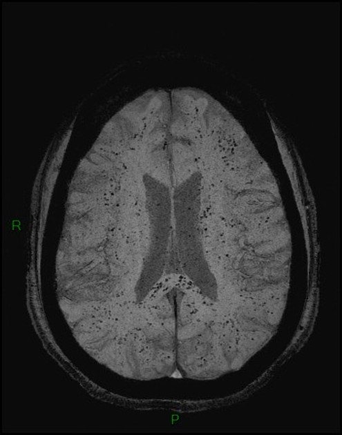
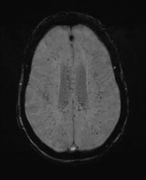
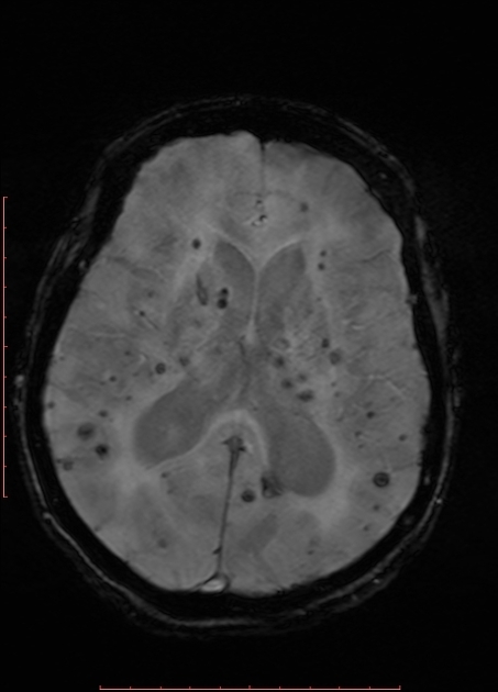
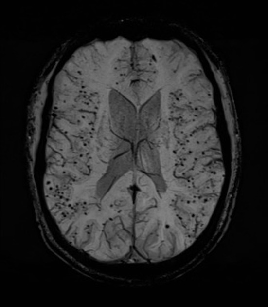
The walnut kernel microbleed pattern along with the starfield pattern and corpus callosum diffusion restriction appears to be the most important imaging markers of cerebral fat embolism 1-3.
In this pattern, there is a diffuse presence of round microbleeds (punctate focal hypointensities) of similar size in the subcortical white matter (involving but not limited to the U-fibers), internal capsule, and the corpus callosum on T2* GRE or, more sensitively, SWI/SWAN images 1. The corona radiata and the non-subcortical centrum semiovale are relatively spared 1. The diffuse involvement of white matter, including outlining the subcortical fibers, resembles a walnut kernel.
Differential diagnosis
Diffuse cerebral microbleeds have been reported in other cases mainly involving the critically ill, such as in patients receiving extracorporeal membrane oxygenation (ECMO), or in critically ill COVID-19 patients, but in these cases, the microhemorrhages appear to be more heterogeneous in size and distribution 4,5.
See also
-
1. Giyab O, Balogh B, Bogner P, Gergely O, Tóth A. Microbleeds Show a Characteristic Distribution in Cerebral Fat Embolism. Insights Imaging. 2021;12(1):42. doi:10.1186/s13244-021-00988-6 - Pubmed
-
2. Kuo K, Pan Y, Lai Y, Cheung W, Chang F, Jarosz J. Dynamic MR Imaging Patterns of Cerebral Fat Embolism: A Systematic Review with Illustrative Cases. AJNR Am J Neuroradiol. 2014;35(6):1052-7. doi:10.3174/ajnr.A3605 - Pubmed
-
3. Parizel P, Demey H, Veeckmans G et al. Early Diagnosis of Cerebral Fat Embolism Syndrome by Diffusion-Weighted MRI (Starfield Pattern). Stroke. 2001;32(12):2942-4. - Pubmed
-
4. Luyt C, Bréchot N, Demondion P et al. Brain Injury During Venovenous Extracorporeal Membrane Oxygenation. Intensive Care Med. 2016;42(5):897-907. doi:10.1007/s00134-016-4318-3 - Pubmed
-
5. Radmanesh A, Derman A, Lui Y et al. COVID-19-Associated Diffuse Leukoencephalopathy and Microhemorrhages. Radiology. 2020;297(1):E223-7. doi:10.1148/radiol.2020202040 - Pubmed
Promoted articles (advertising)


 Unable to process the form. Check for errors and try again.