
Presentation
Ongoing 4th metatarsophalangeal joint pain
Patient Data


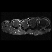

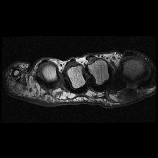
Partial thickness tear of the medial aspect of the 4th toe plantar plate, centered on the medial accessory collateral ligament. The central plantar plate is intact. Small area of fibrosis in the 3rd webspace, likely a pseudo neuroma next to the plantar plate tear.

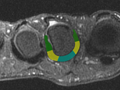
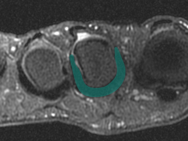
The single color turquoise annotation outlines the intact 2nd toe plantar plate in a horseshoe shape.
The multi-color annotation outlines the different components of an intact plantar plate. The green areas are the proper collateral ligaments (PCL), the yellow the accessory collateral ligaments (ACL), and central, single, and plantar turquoise area the central plantar plate. As there are two PCLs and ACLs, they will be named medial and lateral depending on location, for example, medial accessory collateral ligament. The exact portions of the PCL, ACL, and central plantar plate are not clearly defined on MRI.
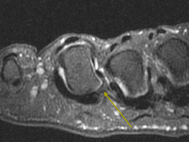
The yellow arrow points to the 4th toe plantar plate tear, which is centered on the medial accessory collateral ligament.
Case Discussion
Plantar plate tears can be partial thickness or full thickness. For full-thickness tears, there must be complete discontinuity of the plantar plate, and there is often angulation of the toe. Knowing the anatomy of the plantar plate will greatly improve MRI interpretation.


 Unable to process the form. Check for errors and try again.