Presentation
Severe headache and dizziness.
Patient Data
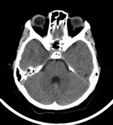


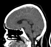

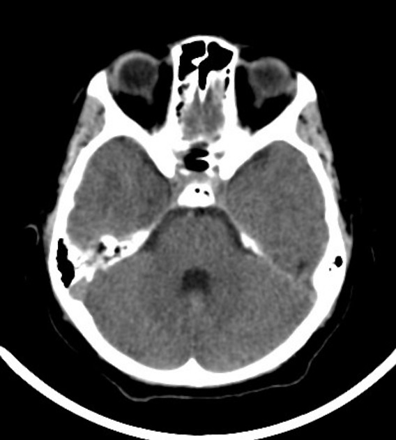
The ventral part of the superior sagittal sinus and its adjacent tributaries are hyperdense, likely superior sagittal sinus and cortical veins thromboses.
The sulci in the right hemisphere and the interhemispheric fissure show linear hyperdensity in keeping with subarachnoid hemorrhage.
Linear hyperdensity in the anterior subcortical part of right parietal lobe suggesting subcortical hemorrhage, mostly due to cortical vein thrombosis.
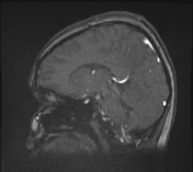

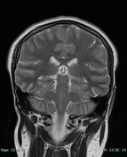

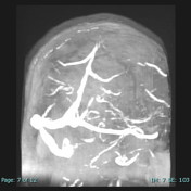



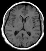

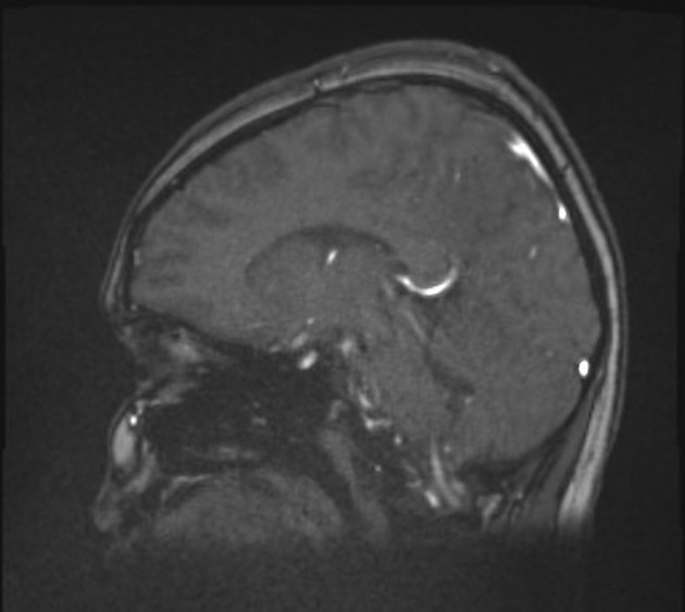
Loss of signal void of ventral part of superior sagittal sinus, along with non-visualization in the MRV study, indicating thrombosed superior sagittal sinus.
Multiple hyperintensities seen in the parasagittal area of the cerebral cortex, cionsistent with cortical veins thromboses.
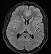
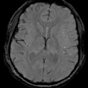
Multiple linear high signal intensities seen in the cortical and interhemispheric sulci in FLAIR images with blooming artifact, representing subarachnoid hemorrhage.
Few foci of intraventricular hemorrhage (blooming artefact) are detected on SWI images in the occipital horns of the lateral ventricles.
Right postcentral subcortical linear hyperintensity on T1 ,T2 and FLAIR images with blooming artifact is seen, in keeping with subacute hemorrhagic focus.
Case Discussion
These findings represent superior sagittal sinus and cortical veins thrombosis complicated by intra-axial, extra-axial and interventricular hemorrhages.


 Unable to process the form. Check for errors and try again.