
Presentation
Intermittent pain in the left gluteal area and limping 6 years after total hip arthroplasty
Patient Data


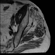
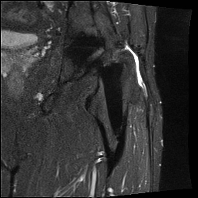
Extensive abductor tendon tear at the greater trochanter: the gluteus minimus tendon and the lateral portion of gluteus medius tendon are retracted, with a gap visible between the greater trochanter and the tendons on the STIR sequence
Trochanteric bursitis with area of high STIR signal intensity lateral to greater trochanter
Fatty muscle degeneration and atrophy of the gluteus minimus and gluteus medius muscle on T1-weighted sequence
Hip arthroplasty: without periprosthetic fracture or adjacent bone marrow edema. However, there is communication between the trochanteric bursitis and the joint effusion.
Case Discussion
Tearing of the abductor tendons and trochanteric bursitis are a common cause of greater trochanteric pain syndrome after total hip arthroplasty (THA). In this case the tendons are retracted and no longer attached to the greater trochanter. There is substantial fatty degeneration of the affected muscles, indicating that this is a chronic condition.
Technical considerations:
In patients with THA metal artifact reduction sequences (MARS) should be used for MRI. The coronal STIR sequence was acquired with the Slice-Encoding for Metal Artifact Correction (SEMAC) technique that addresses both in-plane and through-plane metal artifacts. This sequence allows to detect findings at the bone-metal-interface such as bone marrow edema. Note that in this case the bone-metal-interface is normal. Moreover, the STIR SEMAC sequence is also useful for detection of soft tissue pathology, such as in this case.
The axial T1-weighted sequence was acquired with an increased readout bandwidth, which is a simple but powerful method in order to reduce metal artifacts in non-fat-suppressed images.


 Unable to process the form. Check for errors and try again.