
Presentation
Restricted movements of the left knee.
Patient Data
Age: 40 years
Gender: Male
From the case:
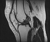
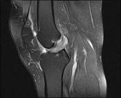
ACL mucoid degeneration - celery stalk sign


Download
Info
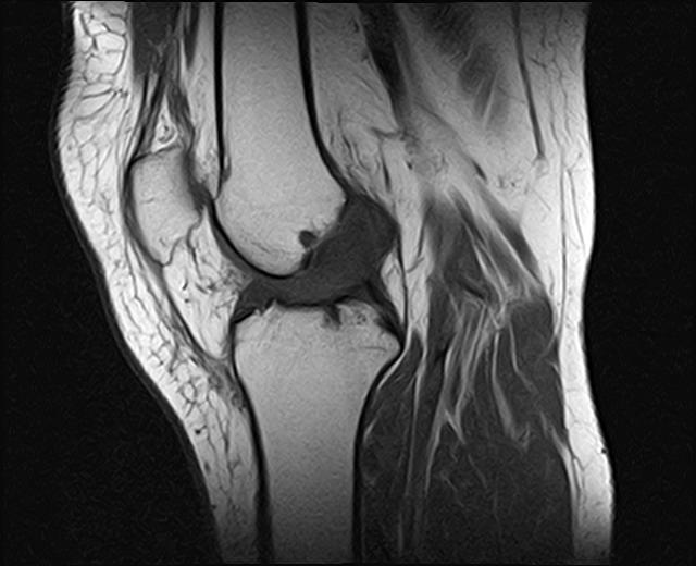
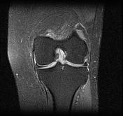
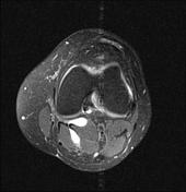
The ACL is thickened, ill-defined of high signal. The longitudinal fibers are intact, separated from each other by higher signal mucinous material well-visualized on T2 and PD fat sat sequences, giving a "celery stalk" appearance.
Mild joint effusion. A small popliteal cyst is noted.
No associated meniscal tear or ligamentous injuries is seen.
Case Discussion
MRI features are most consistent with a mucoid degeneration of the ACL with a "celery stalk sign".


 Unable to process the form. Check for errors and try again.