
Presentation
Headache with dizziness.
Patient Data
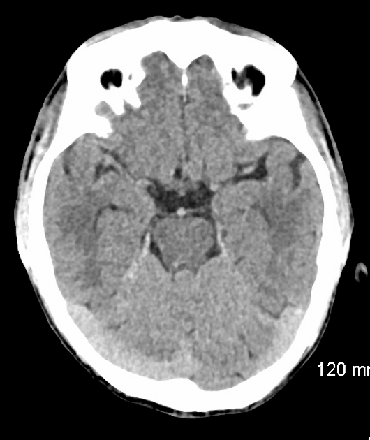
There are acute bilateral cerebellar infarctions (confirmed by diffusion-weighted MRI brain, not shown here). No recent supra-tentorial infarction is seen. No recent intracranial hemorrhage, midline shift, or brain herniation is seen.


Mildly hypoplastic A1 segment of the right anterior cerebral artery.
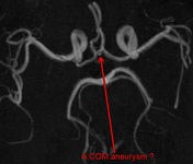
Suspicion of a tiny aneurysm along the anterior communicating artery.
No vascular occlusion or AV malformation is seen.

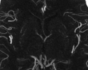
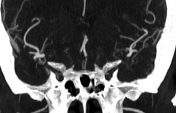
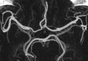
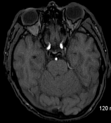
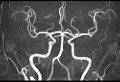
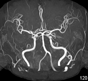
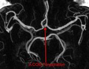
No aneurysm is seen in the circle of Willis, particularly along the anterior communicating artery. No vascular occlusion or AV malformation is seen. Re-demonstration of the mildly hypoplastic A1 segment of the right anterior cerebral artery. Incidental finding of fenestration of the anterior communicating artery and the M1 segment of the right middle cerebral artery.

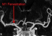

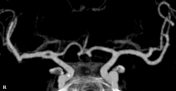
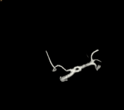
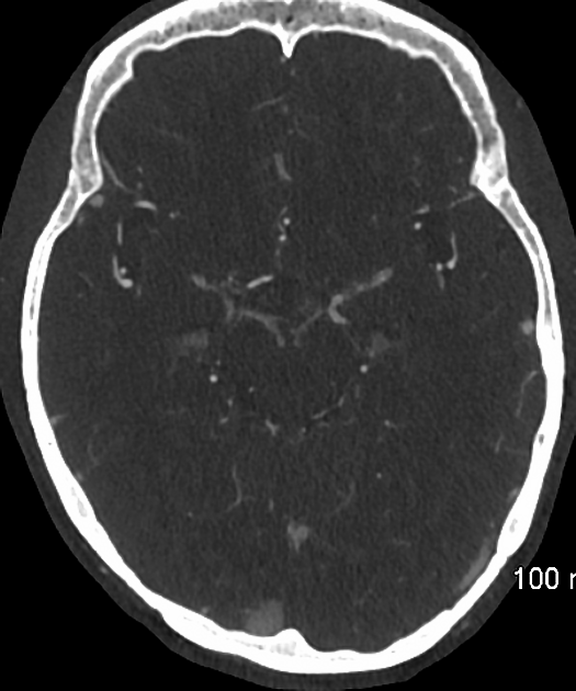
Thick axial and coronal MIP images, along with reformatted VR images, are provided with annotations.
Case Discussion
Fenestration is a normal anatomical variant, seen more commonly in the posterior circulation. Although fenestration has no clinical significance, an association has been observed between fenestration and aneurysm formation. The reported incidence of fenestration in the anterior communicating artery is 5%, and the reported incidence of fenestration in the middle cerebral artery is <1%.


 Unable to process the form. Check for errors and try again.