
Presentation
Past medical history of diabetes mellitus and cholelithiasis presents to ED with the chief complaint of worsening bilateral flank pain, present for the past month.
Patient Data


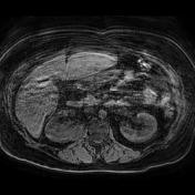
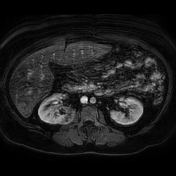
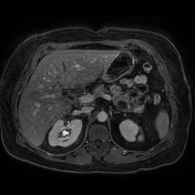
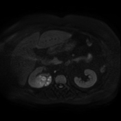
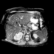
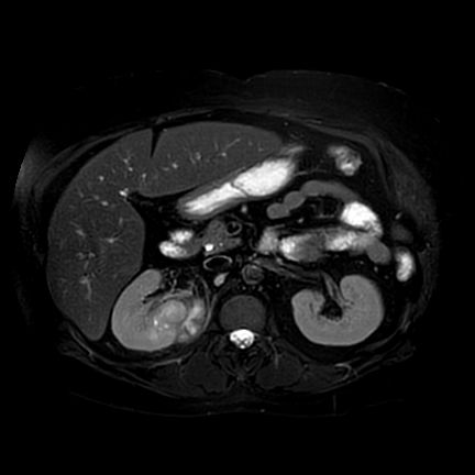
There is a 4.6cm predominantly solid mass within the medial interpolar region of the right kidney. The mass demonstrates mildly increased signal on T2-WI, enhancement, and multifocal small regions of central non-enhancement, cystic change, and/or necrosis. Additionally, the mass demonstrates restricted diffusion. There is signal abnormality and enhancement extending medially to abut the inferomedial right hemidiaphragm and left psoas muscle.
Case Discussion
This patient underwent right nephrectomy for suspected malignancy. The gross pathology report described a "3.3 x 1.5 x 5.0 cm necrotic tan-yellow mass in the lower pole primarily within the corticomedullary parenchyma extending into the renal sinus fat and is adjacent to renal pelvis and calices. The mass extends through the renal capsule into surrounding fat, within 0.2 cm of Gerota’s fascia."
Pathology results returned with a final diagnosis of acute and chronic pyelonephritis with superimposed abscess formation and associated granulomatous inflammation and fat necrosis.


 Unable to process the form. Check for errors and try again.