
Acute left middle cerebral artery territory infarct with clot retrieval
Presentation
Right hemiplegia and aphasia, onset one hour ago.
Patient Data
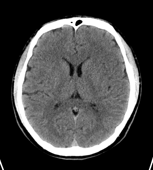
No acute intracranial haemorrhage. Hyperdense left M1 segment. Normal grey-white differentiation.
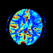

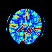
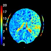
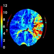
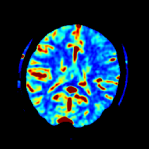
Elevated MTT and Tmax extensively within the left MCA territory. Corresponding areas of reduced CBF but only minor regions of reduced CBV.
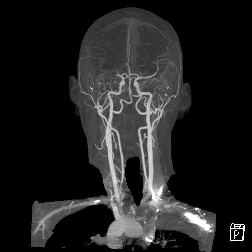
Abrupt truncation of the left M1 extending into M2 branches.

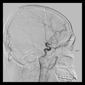
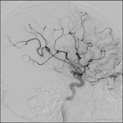
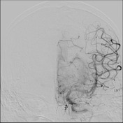
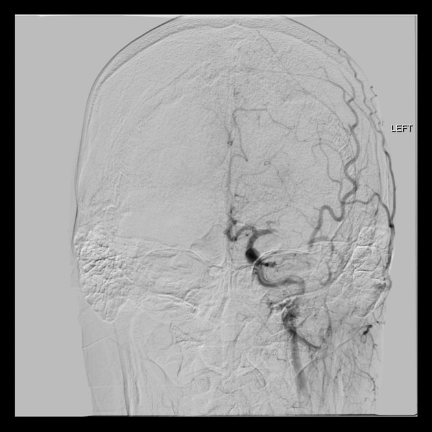
Selective left ICA angiogram confirms a mid M1 occlusion with good collaterals. Two passes with stent retriever with fragmented clot retrieved. TICI 2b. Residual nonocclusive clot within a distal M2 (sylvian) branch.

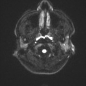
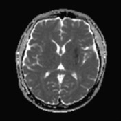
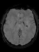
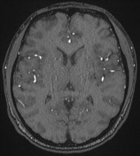

Patchy high T2 signal within the left lentiform nucleus. Anteriorly there is a small area of diffusion restriction. Posteriorly there is blooming artefact consistent with haemorrhage, and diffusion weighted imaging is less reliable. No other evidence of acute stroke or haemorrhage. Old left cerebellar hemisphere infarct. Recanalised left MCA.
Case Discussion
This is an example of the effectiveness of clot retrieval (mechanical thrombectomy) with a potentially large MCA territory stroke being effectively treated resulting in only a very small stroke. The patient had no residual neurological deficit.


 Unable to process the form. Check for errors and try again.