
Presentation
Fever and left ear pus discharge for 15 days.
Patient Data
Age: 10 years
Gender: Female
From the case:
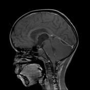
Acute mastoiditis with subdural empyema


Download
Info
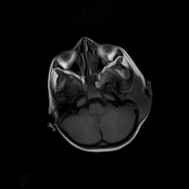
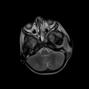
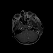
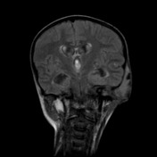
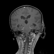
Abnormal signal intensity, low on T1WI , high on T2WI in both mastoid air cells predominantly on the left side, showing extension into the left middle ear cavity. Associated erosion of the lateral wall of mastoid on the left side with juxta-mastoid peripheral rim enhancing fluid collection corresponds to acute coalescent oto-mastoiditis with juxta mastoid abscess.
Bilateral infratentorial subdural abscesses are noted.
Right sided sigmoid sinus thrombus up to the internal jugular vein.
Case Discussion
This case demonstrates acute mastoiditis with intracranial extension demonstrating two complications of severe mastoiditis: subdural empyema and right-sided sigmoid venous thrombus.


 Unable to process the form. Check for errors and try again.