
Presentation
Stepped on a thorn. Penetrating injury to plantar surface of left foot.
Patient Data
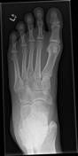

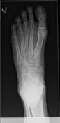
Initial radiograph 3 days after the inciting event is unremarkable.
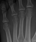

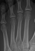
Erosion of the intramedullary trabeculae and cortical destruction of the head of the 4th metatarsal.

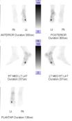
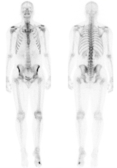
Blood flow and blood pool images show increased radiotracer distribution in the left distal foot, likely localizing to the distal aspect of the 4th metatarsal.
Case Discussion
Acute pyogenic osteomyelitis within the first 48 hours may show subtle soft tissue swelling, loss of fat planes or air within the tissue track from the penetrating injury but most often radiograph is entirely normal. At 1 week the earliest radiograph feature is intramedullary trabeculae destruction. This is followed by enosteal scalloping, cortical destruction and periostitis which is only apparent after 2 weeks. Chronic osteomyelitis is typified by periosteal new bone formation, sequestrum/involucrum which maybe seen from 6 week onwards.


 Unable to process the form. Check for errors and try again.