
Presentation
Profound left optic neuropathy, to rule out a compressive lesion.
Patient Data


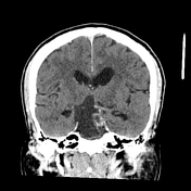
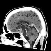
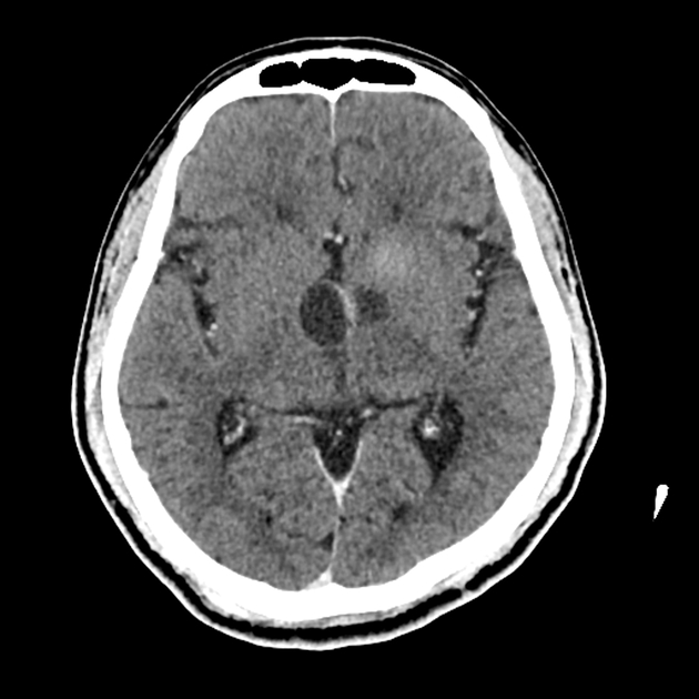
Selected CT images demonstrating a large suprasellar mass composed of cystic and enhancing solid components with scattered tiny calcifications. Significant mass effect on the brainstem and 3rd ventricle with encasement of the basilar artery.

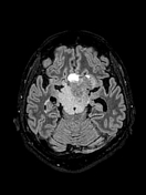
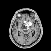
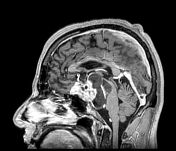
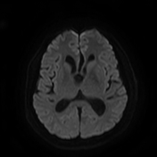
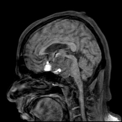
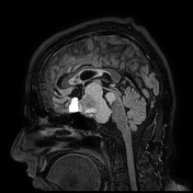
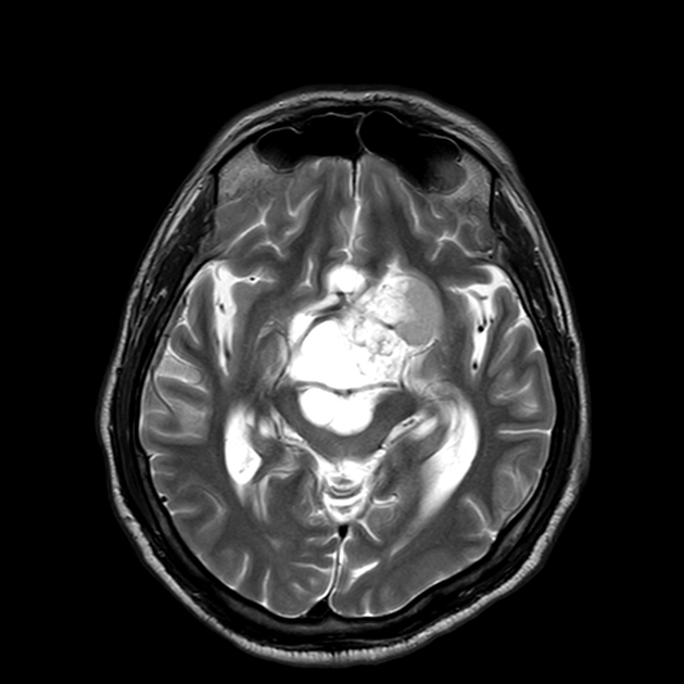
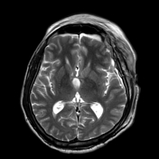
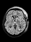
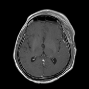
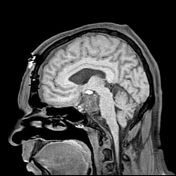
Selected MRI images showing a large suprasellar/hypothalamic mass with cystic and solid enhancing components, resulting in a significant mass effect on the third ventricle, midbrain and the pons. There are also haemorrhagic components.
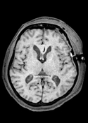

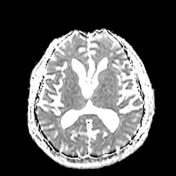
In the interval, a left frontotemporal craniectomy has been carried out with partial resection of the large, suprasellar mass lesion. There is a significant improvement of the mass effect.
Case Discussion
Classic MRI features of an adamantinomatous craniopharyngioma with multiple cysts, some of which are filled with blood products. There are also calcifications, which often occur in the adamantinomatous craniopharyngioma (in about 90% of cases).
In general, adamantinomatous craniopharyngiomas have a bimodal distribution with two peaks (between 5 and 15 years and over 40 years).
The histological report confirmed an adamantinomatous craniopharyngioma (WHO grade 1).


 Unable to process the form. Check for errors and try again.