
Presentation
Heavy menstrual bleeding
Patient Data
Age: 45 years
Gender: Female
From the case:
Adenomyosis


Download
Info
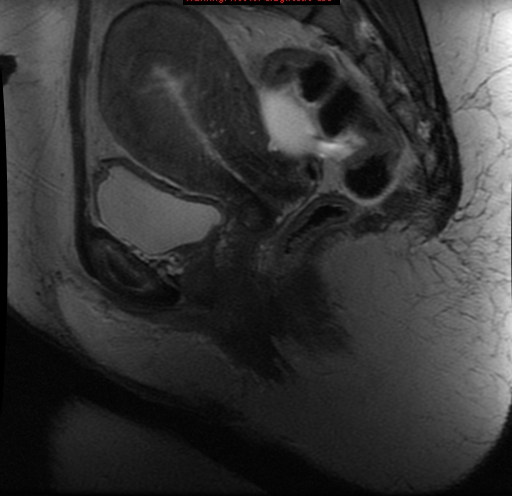
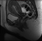
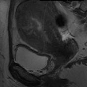
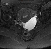
There is an ill-defined ovoid/diffuse region of thickening, with small high T2 signal regions representing small regions of cystic changes. Appearances consistent with adenomyosis.
Case Discussion
MRI demonstrates thickening and blurring of the uterine wall consistent with adenomyosis.


 Unable to process the form. Check for errors and try again.