
Patient Data
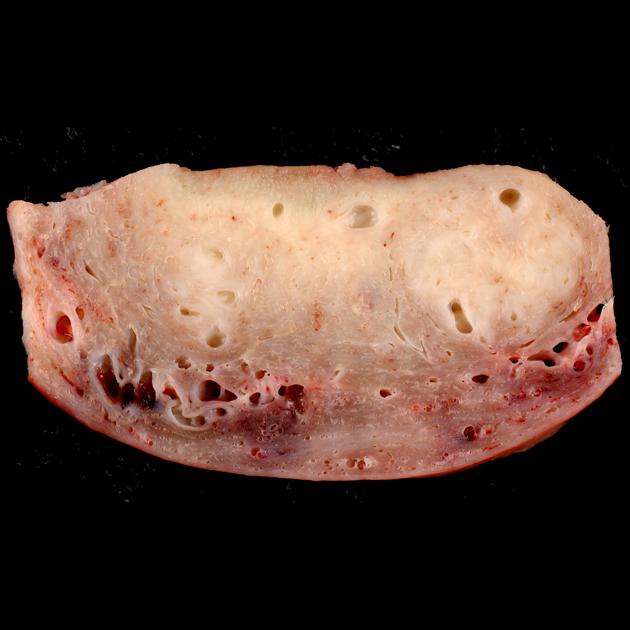
Cross section through the wall of a hysterectomy specimen of a 30-year-old woman who reported chronic pelvic pain and abnormal uterine bleeding. The endometrial surface is at the top of the image, and the serosa is at the bottom. The two pale areas represent regions of adenomyosis.
I think most cases of adenomyosis can be reliably diagnosed grossly by an experienced prosector examining a fixed specimen. Following formalin fixation, the soft adenomyotic areas stand out more strikingly against the firmer myometrium.
Author: Ed Uthman
Original file: wikimedia commons here
License: This file is licensed under the Creative Commons Attribution-Share Alike 2.0 Generic license.
If you believe your copyright or has been infringed please write to license@radiopaedia.org giving details of why you believe this is so.


 Unable to process the form. Check for errors and try again.