
Presentation
Right upper abdominal pain.
Patient Data
Age: 25 years
Gender: Female
From the case:
Adrenal cyst

Show annotations
Download
Info

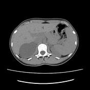
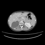
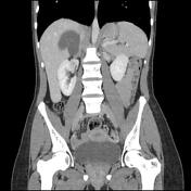
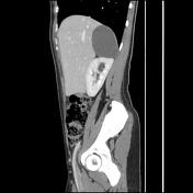
A thin-walled, bi-lobulated right suprarenal cystic mass arising from the adrenal gland with peripheral foci of calcification and homogeneous fluid content. No peripheral enhancement. No solid component or internal septation is seen. A mass effect is noted on segment 7 of the liver and the upper pole of the kidney.
Case Discussion
CT features of an adrenal cyst.
Adrenal cysts are rare lesions with an incidence of <1%. The differential diagnoses include pseudocysts, endothelial cysts, epithelial cysts and parasitic cysts usually hydatid.


 Unable to process the form. Check for errors and try again.