Presentation
Abdominal pain.
Patient Data


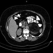
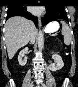
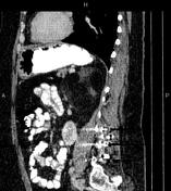
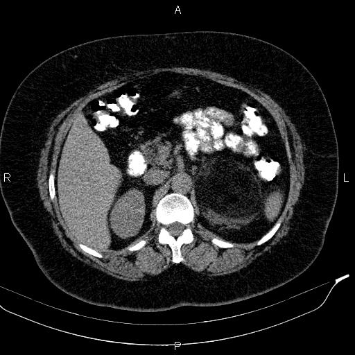
A 95×90×80mm large well defined fat containing mass is noted at anatomical location of left adrenal gland that displaces left kidney inferring. After contrast media administration soft tissue components of the mass shows slightly enhancement.
Post-operative changes are seen due to partial hysterectomy.
Degenerative changes as osteophytosis are seen at the lumbar spine. Additionally, evidence of CD insertion is present at L3, L4 and L5 levels.
Case Discussion
Features on CT scan are most consistent with left adrenal myelolipoma, which is rare, benign and usually asymptomatic tumor of the adrenal gland characterized by the predominance of mature adipocytes.


 Unable to process the form. Check for errors and try again.