
Presentation
Vague abdominal pain.
Patient Data
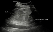
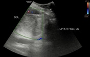
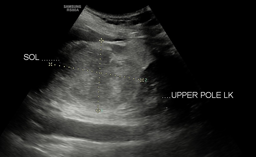
Transverse grayscale and color doppler ultrasound showing a hetero-echoic lesion in the left suprarenal region showing no vascularity on the color doppler study.
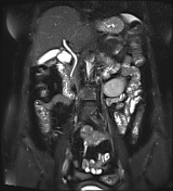

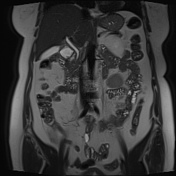
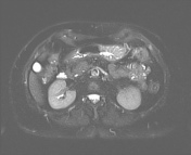
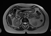
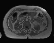
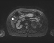


A T1/T2 hyperintense well-defined lesion measuring approx. 7.5x5.3 cm is noted in the left suprarenal region causing compression and inferior displacement of the left kidney. On STIR imaging the lesion appears hypointense suggest fatty nature. Few T1 hypointense septations are noted within the lesion which appears hyperintense on STIR. No restricted diffusion is noted on DWI.
Case Discussion
A T1/T2 hyperintense lesion in the suprarenal region showing suppression on STIR imaging. The left adrenal is not separately visualized from the lesion. Few T1 hypointense septations noted which appear hyperintense on STIR suggest myeloid component. Above mentioned imaging features are almost consistent with myelolipoma.
Other differential diagnoses are retroperitoneal lipoma, well-differentiated liposarcoma, and fat-rich adenoma.


 Unable to process the form. Check for errors and try again.