
Presentation
Incidental finding.
Patient Data
Age: 50 years
Gender: Male
From the case:
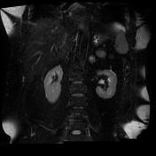
Adrenal myelolipoma


Download
Info
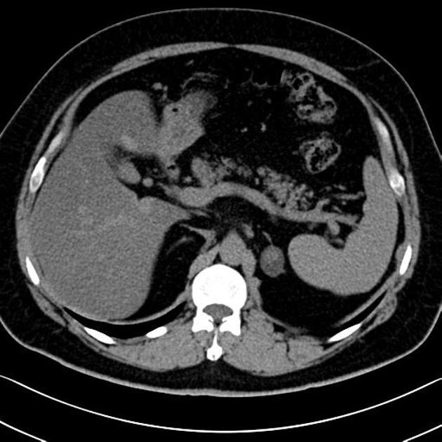
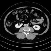
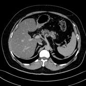
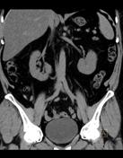
A small, heterogeneous, left adrenal, well-defined lesion is seen. It is primarily of fatty attenuation, with a smaller, soft tissue component centrally. No appreciable enhancement can be seen. Features consistent with a left adrenal myelolipoma.
No retroperitoneal hemorrhage.
Diffuse hepatic steatosis.
No further abnormality is seen in the abdominal solid viscera.
From the case:
Adrenal myelolipoma


Download
Info
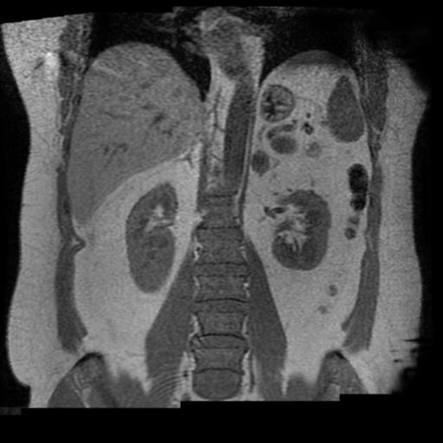
The out-of-phase images show striking drop of signal intensity, due to the lesion comprising an admixture of soft tissue and fat.
Case Discussion
Adrenal myelolipoma


 Unable to process the form. Check for errors and try again.