
Patient Data
Gender: Male
Note: This case has been tagged as "legacy" as it no longer meets image preparation and/or other case publication guidelines.
From the case:
Agenesis of the corpus callosum

Download
Info
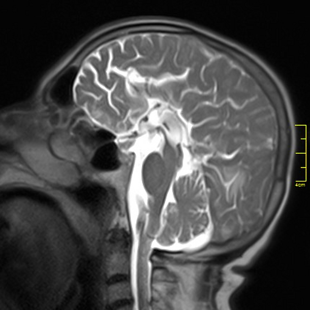
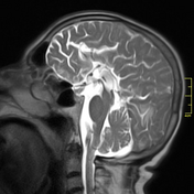
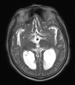
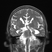
The corpus callosum is absent.
Axial images: The bodies of lateral ventricles have a parallel orientation. Fibers of white matter located in the medial side of bodies of lateral ventricles are called "Probst bundles". These are considered as the fibers that meant to make corpus callosum. The posterior parts of lateral ventricles are enlarged; this is called "colpocephaly".
Coronal images: The frontal horns make "steer horn", and "Viking helmet" signs of the lateral ventricles in the coronal plane.
Sagittal image: Brain sulci have a "sun ray" appearance in the midsagittal plane.


 Unable to process the form. Check for errors and try again.